
12 months from date of receipt / reconstitution, -20°C as supplied
| 应用 | 稀释度 |
|---|---|
| WB | 1:1000 |
| IHC | 1:2000 |
| ICC | 1:50 |
Soluble transferrin receptor conventionally refers to the cleaved extracellular portion of transferrin receptor 1 that is released into serum. This receptor is a protein dimer of two identical subunits, linked together by two pairs of disulfide bonds. Its molecular mass 190,000 Dalton. Blood testing of the soluble transferrin receptor (sTfR) is used as a measure of functional iron status and the investigation of iron deficiency anemia. Ferritin, a routine investigation for anemia, is an acute-phase reactant, and may be elevated in states of inflammation, thereby falsely indicating that iron stores are adequate. Because sTfR is insensitive to inflammation, it can detect anemia in patients with preexisting inflammatory states, and is particularly useful in distinguishing between the anemia of chronic disease and anemias caused by lack of iron intake.
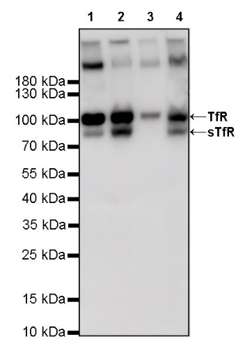
WB result of sTfR Rabbit mAb
Primary antibody: sTfR Rabbit mAb at 1/1000 dilution
Lane 1: HeLa whole cell lysate 20 µg
Lane 2: K562 whole cell lysate 20 µg
Lane 3: HT-1080 whole cell lysate 20 µg
Lane 4: 293T whole cell lysate 20 µg
Secondary antibody: Goat Anti-Rabbit IgG, (H+L), HRP conjugated at 1/10000 dilution
Predicted MW: 84 kDa
Observed MW: 100, 85 kDa
IHC shows positive staining in paraffin-embedded human kidney. Anti-sTfR antibody was used at 1/2000 dilution, followed by a HRP Polymer for Mouse & Rabbit IgG (ready to use). Counterstained with hematoxylin. Heat mediated antigen retrieval with Tris/EDTA buffer pH9.0 was performed before commencing with IHC staining protocol.
IHC shows positive staining in paraffin-embedded human liver. Anti-sTfR antibody was used at 1/2000 dilution, followed by a HRP Polymer for Mouse & Rabbit IgG (ready to use). Counterstained with hematoxylin. Heat mediated antigen retrieval with Tris/EDTA buffer pH9.0 was performed before commencing with IHC staining protocol.
IHC shows positive staining in paraffin-embedded human placenta. Anti-sTfR antibody was used at 1/2000 dilution, followed by a HRP Polymer for Mouse & Rabbit IgG (ready to use). Counterstained with hematoxylin. Heat mediated antigen retrieval with Tris/EDTA buffer pH9.0 was performed before commencing with IHC staining protocol.
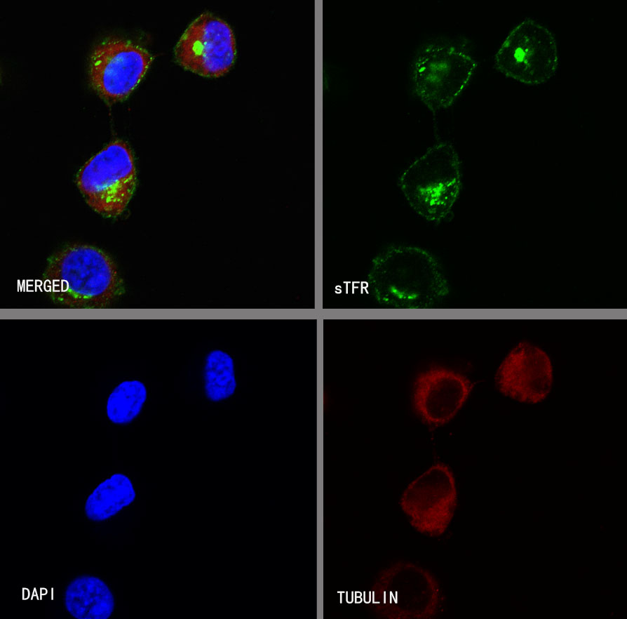
ICC shows clustered cytoplasm staining in HT-1080 cells treated with BFA (5 μg/ml) for 15min. Anti-sTfR antibody was used at 1/50 dilution (Green) and incubated overnight at 4°C. Goat polyclonal Antibody to Rabbit IgG - H&L (Alexa Fluor® 488) was used as secondary antibody at 1/1000 dilution. The cells were fixed with 100% ice-cold methanol and permeabilized with 0.1% PBS-Triton X-100. Nuclei were counterstained with DAPI (Blue). Counterstain with tubulin (Red).
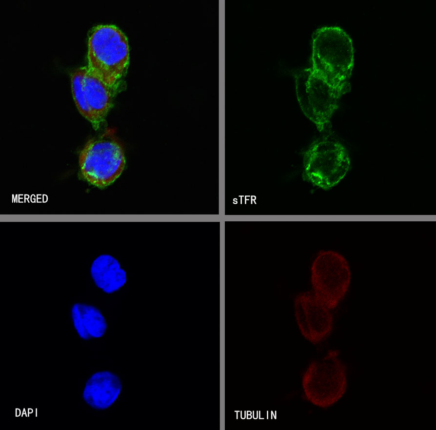
Negative control: ICC shows homogeneous cytoplasm staining in HT-1080 cells untreated with BFA (5 μg/ml) for 15min. Anti-sTfR antibody was used at 1/50 dilution (Green) and incubated overnight at 4°C. Goat polyclonal Antibody to Rabbit IgG - H&L (Alexa Fluor® 488) was used as secondary antibody at 1/1000 dilution. The cells were fixed with 100% ice-cold methanol and permeabilized with 0.1% PBS-Triton X-100. Nuclei were counterstained with DAPI (Blue). Counterstain with tubulin (Red).
 您现在的位置: