
12 months from date of receipt / reconstitution, -20 °C as supplied
| 应用 | 稀释度 |
|---|---|
| WB | 1:1000 |
| IHC | 1:500-1:2000 |
Protein NDRG1 is a protein that in humans is encoded by the NDRG1 gene. This gene is a member of the N-myc downregulated gene family which belongs to the alpha/beta hydrolase superfamily. The protein encoded by this gene is a cytoplasmic protein involved in stress responses, hormone responses, cell growth, and differentiation. Mutations in this gene have been reported to be causative the autosomal-recessive version of Charcot-Marie-Tooth disease known as CMT4D. NDRG1 is a potent, iron-regulated growth and metastasis suppressor that was found to be negatively correlated with cancer progression in a number of tumors, including prostate, pancreatic, breast, and colon cancers. NDRG1 has marked anti-oncogenic activity, being associated with decreased cell proliferation, migration, invasion, and angiogenesis. The molecular functions of NDRG1 affect numerous signaling pathways that regulate cancer cell proliferation, invasion, angiogenesis, and migration.
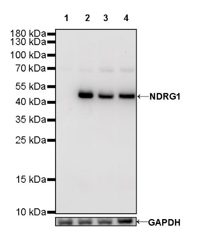
WB result of NDRG1 Rabbit mAb
Primary antibody: NDRG1 Rabbit mAb at 1/1000 dilution
Lane 1: PANC-1 whole cell lysate 20 µg
Lane 2: PC-3 whole cell lysate 20 µg
Lane 3: HeLa whole cell lysate 20 µg
Lane 4: Jurkat whole cell lysate 20 µg
Negative control: PANC-1 whole cell lysate
Secondary antibody: Goat Anti-Rabbit IgG, (H+L), HRP conjugated at 1/10000 dilution
Predicted MW: 43 kDa
Observed MW: 48 kDa
(This blot was developed with high sensitivity substrate)
WB result of NDRG1 Rabbit mAb
Primary antibody: NDRG1 Rabbit mAb at 1/1000 dilution
Lane 1: mouse brain lysate 20 µg
Secondary antibody: Goat Anti-Rabbit IgG, (H+L), HRP conjugated at 1/10000 dilution
Predicted MW: 43 kDa
Observed MW: 48 kDa
(This blot was developed with high sensitivity substrate)
WB result of NDRG1 Rabbit mAb
Primary antibody: NDRG1 Rabbit mAb at 1/1000 dilution
Lane 1: rat brain lysate 20 µg
Secondary antibody: Goat Anti-Rabbit IgG, (H+L), HRP conjugated at 1/10000 dilution
Predicted MW: 43 kDa
Observed MW: 48 kDa
(This blot was developed with high sensitivity substrate)
IHC shows positive staining in paraffin-embedded human kidney. Anti-NDRG1 antibody was used at 1/2000 dilution, followed by a HRP Polymer for Mouse & Rabbit IgG (ready to use). Counterstained with hematoxylin. Heat mediated antigen retrieval with Tris/EDTA buffer pH9.0 was performed before commencing with IHC staining protocol.
IHC shows positive staining in paraffin-embedded human prostate. Anti-NDRG1 antibody was used at 1/2000 dilution, followed by a HRP Polymer for Mouse & Rabbit IgG (ready to use). Counterstained with hematoxylin. Heat mediated antigen retrieval with Tris/EDTA buffer pH9.0 was performed before commencing with IHC staining protocol.
IHC shows positive staining in paraffin-embedded human colon cancer. Anti-NDRG1 antibody was used at 1/2000 dilution, followed by a HRP Polymer for Mouse & Rabbit IgG (ready to use). Counterstained with hematoxylin. Heat mediated antigen retrieval with Tris/EDTA buffer pH9.0 was performed before commencing with IHC staining protocol.
IHC shows positive staining in paraffin-embedded human hepatocellular carcinoma. Anti-NDRG1 antibody was used at 1/2000 dilution, followed by a HRP Polymer for Mouse & Rabbit IgG (ready to use). Counterstained with hematoxylin. Heat mediated antigen retrieval with Tris/EDTA buffer pH9.0 was performed before commencing with IHC staining protocol.
IHC shows positive staining in paraffin-embedded mouse colon. Anti-NDRG1 antibody was used at 1/500 dilution, followed by a HRP Polymer for Mouse & Rabbit IgG (ready to use). Counterstained with hematoxylin. Heat mediated antigen retrieval with Tris/EDTA buffer pH9.0 was performed before commencing with IHC staining protocol.
IHC shows positive staining in paraffin-embedded rat colon. Anti-NDRG1 antibody was used at 1/500 dilution, followed by a HRP Polymer for Mouse & Rabbit IgG (ready to use). Counterstained with hematoxylin. Heat mediated antigen retrieval with Tris/EDTA buffer pH9.0 was performed before commencing with IHC staining protocol.
 您现在的位置: