Protein sequence (P06332, Lys27-Thr394, with C-10*His) KTLVLGKEGESAELPCESSQKKITVFTWKFSDQRKILGQHGKGVLIRGGSPSQFDRFDSKKGAWEKGSFPLIINKLKMEDSQTYICELENRKEEVELWVFKVTFSPGTSLLQGQSLTLTLDSNSKVSNPLTECKHKKGKVVSGSKVLSMSNLRVQDSDFWNCTVTLDQKKNWFGMTLSVLGFQSTAITAYKSEGESAEFSFPLNFAEENGWGELMWKAEKDSFFQPWISFSIKNKEVSVQKSTKDLKLQLKETLPLTLKIPQVSLQFAGSGNLTLTLDKGTLHQEVNLVVMKVAQLNNTLTCEVMGPTSPKMRLTLKQENQEARVSEEQKVVQVVAPETGLWQCLLSEGDKVKMDSRIQVLSRGVNQTGGGGSHHHHHHHHHH
12 months from date of receipt, -20 to -70 °C as supplied. 6 months, -20 to -70 °C under sterile conditions after reconstitution. 1 week, 2 to 8 °C under sterile conditions after reconstitution. Please avoid repeated freeze-thaw cycles.
In molecular biology, CD4 (cluster of differentiation 4) is a glycoprotein that serves as a co-receptor for the T-cell receptor (TCR). CD4 is found on the surface of immune cells such as T helper cells, monocytes, macrophages, and dendritic cells. CD4+ T helper cells are white blood cells that are an essential part of the human immune system. CD4 continues to be expressed in most neoplasms derived from T helper cells. It is therefore possible to use CD4 immunohistochemistry on tissue biopsy samples to identify most forms of peripheral T cell lymphoma and related malignant conditions. The antigen has also been associated with a number of autoimmune diseases such as vitiligo and type I diabetes mellitus. T-cells play a large part in autoinflammatory diseases. When testing a drug's efficacy or studying diseases, it is helpful to quantify the amount of T-cells on fresh-frozen tissue with CD4+, CD8+, and CD3+ T-cell markers.
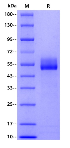
2μg(R: reducing conditions)
 您现在的位置: