
12 months from date of receipt / reconstitution, -20 °C as supplied
| 应用 | 稀释度 |
|---|---|
| WB | 1:1000 |
| IP | 1:50 |
| IHC | 1:500 |
| ICC | 1:500 |
Tyrosine phosphorylation is the addition of a phosphate (PO43−) group to the amino acid tyrosine on a protein. It is one of the main types of protein phosphorylation. This transfer is made possible through enzymes called tyrosine kinases. Tyrosine phosphorylation is a key step in signal transduction and the regulation of enzymatic activity.
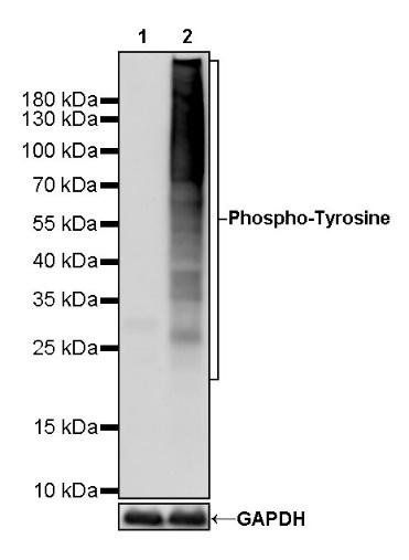
WB result of Phospho-Tyrosine Rabbit mAb
Primary antibody: Phospho-Tyrosine Rabbit mAb at 1/1000 dilution
Lane 1: HeLa whole cell lysate 20 µg
Lane 2: HeLa treated with Sodium pervanadate (3 mM, 30 min) whole cell lysate 20 µg
Secondary antibody: Goat Anti-Rabbit IgG, (H+L), HRP conjugated at 1/10000 dilution
Predicted MW: multiple
Observed MW: multiple
Phospho-Tyrosine Rabbit mAb at 1/50 dilution (1 µg) immunoprecipitating Phospho-Tyrosine in 0.4 mg HeLa treated with Sodium pervanadate (3mM, 30min) whole cell lysate.
Western blot was performed on the immunoprecipitate using Phospho-Tyrosine Rabbit mAb at 1/1000 dilution.
Secondary antibody (HRP) for IP was used at 1/400 dilution.
Lane 1: HeLa treated with Sodium pervanadate (3 mM, 30 min) whole cell lysate 20 µg (Input)
Lane 2: Phospho-Tyrosine Rabbit mAb IP in HeLa treated with Sodium pervanadate (3mM, 30min) whole cell lysate
Lane 3: Rabbit monoclonal IgG IP in HeLa treated with Sodium pervanadate (3mM, 30min) whole cell lysate
Predicted MW: multiple
Observed MW: multiple
IHC shows positive staining in paraffin-embedded mouse lung (left) and negative staining in phosphatase treated (37 °C, 2h) paraffin-embedded mouse lung (right). Anti- Phospho-Tyrosine antibody was used at 1/500 dilution, followed by a HRP Polymer for Mouse & Rabbit IgG (ready to use). Counterstained with hematoxylin. Heat mediated antigen retrieval with Tris/EDTA buffer pH9.0 was performed before commencing with IHC staining protocol.
IHC shows positive staining in paraffin-embedded rat lung (left) and negative staining in phosphatase treated (37 °C, 2h) paraffin-embedded mouse lung (right). Anti- Phospho-Tyrosine antibody was used at 1/500 dilution, followed by a HRP Polymer for Mouse & Rabbit IgG (ready to use). Counterstained with hematoxylin. Heat mediated antigen retrieval with Tris/EDTA buffer pH9.0 was performed before commencing with IHC staining protocol.
IHC shows positive staining in paraffin-embedded rat stomach (left) and negative staining in phosphatase treated (37 °C, 2h) paraffin-embedded mouse lung (right). Anti- Phospho-Tyrosine antibody was used at 1/500 dilution, followed by a HRP Polymer for Mouse & Rabbit IgG (ready to use). Counterstained with hematoxylin. Heat mediated antigen retrieval with Tris/EDTA buffer pH9.0 was performed before commencing with IHC staining protocol.
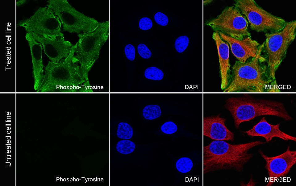
ICC analysis of HeLa cells treated with Sodium pervanadate (3mM, 30min) (top panel) and HeLa cells untreated with Sodium pervanadate (3mM, 30min) (below panel). Anti-Phospho-Tyrosine antibody was used at 1/500 dilution (Green) and incubated overnight at 4°C. Goat polyclonal Antibody to Rabbit IgG - H&L (Alexa Fluor® 488) was used as secondary antibody at 1/1000 dilution. The cells were fixed with 4% PFA and permeabilized with 0.1% PBS-Triton X-100. Nuclei were counterstained with DAPI (Blue). Counterstain with tubulin (Red).
 您现在的位置: