
12 months from date of receipt / reconstitution, -20 °C as supplied
| 应用 | 稀释度 |
|---|---|
| WB | 1:500 |
| IP | 1:25 |
| IHC | 1:1000-1:2000 |
Synucleins are a family of soluble proteins common to vertebrates, primarily expressed in neural tissue and in certain tumors. The synuclein family includes three known proteins: alpha-synuclein, beta-synuclein, and gamma-synuclein. Interest in the synuclein family began when alpha-synuclein was found to be mutated in several families with autosomal dominant Parkinson's disease. All synucleins have in common a highly conserved alpha-helical lipid-binding motif with similarity to the class-A2 lipid-binding domains of the exchangeable apolipoproteins. Normal cellular functions have not been determined for any of the synuclein proteins. Some data suggest a role in the regulation of membrane stability and/or turnover. Mutations in alpha-synuclein are associated with early-onset familial Parkinson's disease and the protein aggregates abnormally in Parkinson's disease, Lewy body disease, and other neurodegenerative diseases. Beta-synuclein is suggested to be an inhibitor of alpha-synuclein aggregation, which occurs in neurodegenerative diseases such as Parkinson's disease. Thus, beta-synuclein may protect the central nervous system from the neurotoxic effects of alpha-synuclein and provide a novel treatment of neurodegenerative disorders.
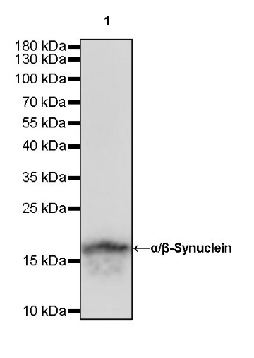
WB result of α/β-Synuclein Rabbit mAb
Primary antibody: α/β-Synuclein Rabbit mAb at 1/500 dilution
Lane 1: mouse brain lysate 20 µg
Secondary antibody: Goat Anti-Rabbit IgG, (H+L), HRP conjugated at 1/10000 dilution
Predicted MW: 14 kDa
Observed MW: 18 kDa
WB result of α/β-Synuclein Rabbit mAb
Primary antibody: α/β-Synuclein Rabbit mAb at 1/500 dilution
Lane 1: rat brain lysate 20 µg
Secondary antibody: Goat Anti-Rabbit IgG, (H+L), HRP conjugated at 1/10000 dilution
Predicted MW: 14 kDa
Observed MW: 18 kDa
α/β-Synuclein Rabbit mAb at 1/25 dilution (1 µg) immunoprecipitating α/β-Synuclein in 0.4 mg mouse brain lysate.
Western blot was performed on the immunoprecipitate using α/β-Synuclein Rabbit mAb at 1/500 dilution.
Secondary antibody (HRP) for IP was used at 1/400 dilution.
Lane 1: mouse brain lysate 20 µg (Input)
Lane 2: α/β-Synuclein Rabbit mAb IP in mouse brain lysate
Lane 3: Rabbit monoclonal IgG IP in mouse brain lysate
Predicted MW: 14 kDa
Observed MW: 18 kDa
IHC shows positive staining in paraffin-embedded human cerebral cortex. Anti-α/β-Synuclein antibody was used at 1/1000 dilution, followed by a HRP Polymer for Mouse & Rabbit IgG (ready to use). Counterstained with hematoxylin. Heat mediated antigen retrieval with Tris/EDTA buffer pH9.0 was performed before commencing with IHC staining protocol.
Negative control: IHC shows negative staining in paraffin-embedded human colon. Anti-α/β-Synuclein antibody was used at 1/1000 dilution, followed by a HRP Polymer for Mouse & Rabbit IgG (ready to use). Counterstained with hematoxylin. Heat mediated antigen retrieval with Tris/EDTA buffer pH9.0 was performed before commencing with IHC staining protocol.
Negative control: IHC shows negative staining in paraffin-embedded human liver. Anti-α/β-Synuclein antibody was used at 1/1000 dilution, followed by a HRP Polymer for Mouse & Rabbit IgG (ready to use). Counterstained with hematoxylin. Heat mediated antigen retrieval with Tris/EDTA buffer pH9.0 was performed before commencing with IHC staining protocol.
Negative control: IHC shows negative staining in paraffin-embedded human tonsil. Anti-α/β-Synuclein antibody was used at 1/1000 dilution, followed by a HRP Polymer for Mouse & Rabbit IgG (ready to use). Counterstained with hematoxylin. Heat mediated antigen retrieval with Tris/EDTA buffer pH9.0 was performed before commencing with IHC staining protocol.
Negative control: IHC shows negative staining in paraffin-embedded human cervical squamous cell carcinoma. Anti-α/β-Synuclein antibody was used at 1/1000 dilution, followed by a HRP Polymer for Mouse & Rabbit IgG (ready to use). Counterstained with hematoxylin. Heat mediated antigen retrieval with Tris/EDTA buffer pH9.0 was performed before commencing with IHC staining protocol.
Negative control: IHC shows negative staining in paraffin-embedded human endometrial carcinoma. Anti-α/β-Synuclein antibody was used at 1/1000 dilution, followed by a HRP Polymer for Mouse & Rabbit IgG (ready to use). Counterstained with hematoxylin. Heat mediated antigen retrieval with Tris/EDTA buffer pH9.0 was performed before commencing with IHC staining protocol.
IHC shows positive staining in paraffin-embedded mouse cerebral cortex. Anti-α/β-Synuclein antibody was used at 1/1000 dilution, followed by a HRP Polymer for Mouse & Rabbit IgG (ready to use). Counterstained with hematoxylin. Heat mediated antigen retrieval with Tris/EDTA buffer pH9.0 was performed before commencing with IHC staining protocol.
IHC shows positive staining in paraffin-embedded rat cerebral cortex. Anti-α/β-Synuclein antibody was used at 1/1000 dilution, followed by a HRP Polymer for Mouse & Rabbit IgG (ready to use). Counterstained with hematoxylin. Heat mediated antigen retrieval with Tris/EDTA buffer pH9.0 was performed before commencing with IHC staining protocol.
 您现在的位置: