
PBS, 40% Glycerol, 0.05% BSA, 0.03% Proclin 300
12 months from date of receipt / reconstitution, -20 °C as supplied
| 应用 | 稀释度 |
|---|---|
| WB | 1:1000 |
| IP | 1:50 |
| ICC | 1:500 |
| ICFCM | 1:5000 |
| ChIP | 1:20-1:50 |
The green fluorescent protein (GFP) is a protein that exhibits bright green fluorescence when exposed to light in the blue to ultraviolet range.[PMID: 28749] The label GFP traditionally refers to the protein first isolated from the jellyfish Aequorea victoria and is sometimes called avGFP.
WB result of GFP Rabbit mAb
Primary antibody: GFP Rabbit mAb at 1/1000 dilution
Lane 1: 293T transfected with GFP whole cell lysate 5 µg
Secondary antibody: Goat Anti-Rabbit IgG, (H+L), HRP conjugated at 1/10000 dilution
Predicted MW: 27 kDa
Observed MW: 32 kDa
Exposure time: 30 s
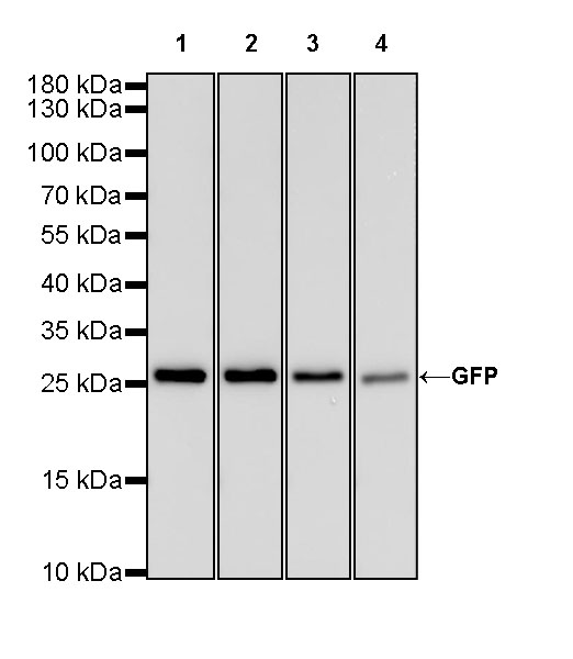
WB result of GFP Rabbit mAb
Lysate: Aequorea victoria GFP Protein 20ng
Primary antibody:
Lane 1: GFP Rabbit mAb at 1/5000 dilution
Lane 2: GFP Rabbit mAb at 1/10000 dilution
Lane 3: GFP Rabbit mAb at 1/50000 dilution
Lane 4: GFP Rabbit mAb at 1/100000 dilution
Secondary antibody: Goat Anti-Rabbit IgG, (H+L), HRP conjugated at 1/10000 dilution
Predicted MW: 28 kDa
Observed MW: 28 kDa
Flow cytometric analysis of 4% PAF fixed 90% methanol permeabilized GFP-transfected 293T cells labelling GFP antibody at 1/5000 (0.01 μg) dilution / (right panel) compared with a Rabbit IgG Isotype Control / (left panel). Goat Anti-Rabbit IgG Alexa Fluor 647 was used as the secondary antibody.
GFP Rabbit mAb at 1/50 dilution (0.5 µg) immunoprecipitating GFP in 0.2 mg 293T transfected with GFP whole cell lysate.
Western blot was performed on the immunoprecipitate using GFP Rabbit mAb at 1/1000 dilution.
Secondary antibody (HRP) for IP was used at 1/400 dilution.
Lane 1: 293T transfected with GFP whole cell lysate 5 µg (Input)
Lane 2: GFP Rabbit mAb IP in 293T transfected with GFP whole cell lysate
Lane 3: Rabbit monoclonal IgG IP in 293T transfected with GFP whole cell lysate
Predicted MW: 27 kDa
Observed MW: 27 kDa
ICC shows positive staining in GFP-transfected HeLa cells. Anti-GFP antibody was used at 1/500 dilution (Red) and incubated overnight at 4°C. Goat polyclonal Antibody to Rabbit IgG - H&L (Alexa Fluor® 594) was used as secondary antibody at 1/1000 dilution. The cells were fixed with 4% PFA and permeabilized with 0.1% PBS-Triton X-100. Nuclei were counterstained with DAPI (Blue).
Negative control: ICC shows negative staining in Vector-transfected HeLa cells. Anti-GFP antibody was used at 1/500 dilution and incubated overnight at 4°C. Goat polyclonal Antibody to Rabbit IgG - H&L (Alexa Fluor® 594) was used as secondary antibody at 1/1000 dilution. The cells were fixed with 4% PFA and permeabilized with 0.1% PBS-Triton X-100. Nuclei were counterstained with DAPI (Blue).
Chromatin immunoprecipitation (ChIP) was
performed on 293T cells were either untransfected
(left panel) or transfected with an GFP-tagged human
H3 construct (right panel) cross - linked with 1%
formaldehyde for 10 min, then chromatin was
fragmented by sonication.
Parallel reactions used GFP Recombinant Rabbit mAb
(S-296-169), Histone H3 Recombinant Rabbit mAb
(SDT-266-44) and Rabbit mAb IgG Isotype
Control (SDT-R173) at 1:50 for immunoprecipitation.
Post - immunoprecipitation, both samples were washed,
eluted, and cross - links reversed. Purified DNA was
analyzed by qPCR.
qPCR (%input: immunoprecipitated DNA/input DNA)
showed the enrichment of RPL30, MYOD1 and SAT-2 in
GFP Recombinant Rabbit mAb (S-296-169)-
immunoprecipitated sample.
 您现在的位置: