
| 应用 | 稀释度 |
|---|---|
| WB | 1:1000 |
| IHC-P | 1:1000-1:2000 |
| IP | 1:50 |
Cluster of differentiation 146 (CD146) is a cell adhesion molecule (CAM) which belongs to the immunoglobulin superfamily (IgSF) [PMID: 3542195]. Human CD146 has previously been designated several synonyms, including MUC18 [PMID: 3542195, PMID: 2602381], A32 antigen [PMID: 8162602,PMID: 7943174], S-Endo-1 [PMID: 8573133], melanoma CAM (MCAM or Mel-CAM) [PMID: 7943174, PMID: 8616875, PMID: 9187135], metastasis CAM (MET-CAM) and hemopoietic CAM (HEMCAM). The avian homolog of CD146 has been named gicerin [PMID: 8161457]. CD146 was originally identified as a marker for melanoma (MCAM), due to its overexpression in metastatic lesions and advanced primary tumors, yet not in benign lesions. Increasing amounts of evidence have demonstrated that CD146 is overexpressed in a variety of carcinomas, in addition to melanomas [PMID: 16804906, PMID: 22826148, PMID: 22754372, PMID: 22210108]. As a result of this characteristic, CD146 has attracted attention and is considered to be a potential marker for tumor diagnosis, prognosis and treatment. The majority of studies support the theory that CD146 promotes tumor growth, angiogenesis and metastasis [PMID: 19356677], therefore, CD146 is a promising target for tumor therapy [PMID: 17121934, PMID: 22977083].

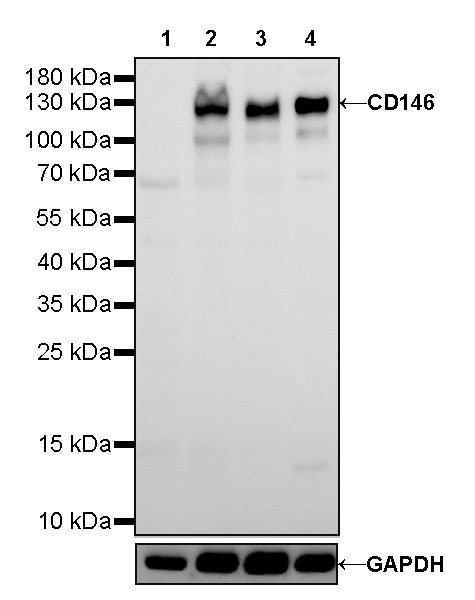
WB result of CD146 Rabbit mAb
Primary antibody: CD146 Rabbit mAb at 1/1000 dilution
Lane 1: LNCaP whole cell lysate 20 µg
Lane 2: SK-MEL-28 whole cell lysate 20 µg
Lane 3: HeLa whole cell lysate 20 µg
Lane 4: HUVEC whole cell lysate 20 µg
Negative control: LNCaP whole cell lysate
Secondary antibody: Goat Anti-Rabbit IgG, (H+L), HRP conjugated at 1/10000 dilution
Predicted MW: 72kDa
Observed MW: 120kDa

CD146 Rabbit mAb at 1/50 dilution (1 µg) immunoprecipitating CD146 in 0.4 mg HeLa whole cell lysate.
Western blot was performed on the immunoprecipitate using CD146 Rabbit mAb at 1/1000 dilution.
Secondary antibody (HRP) for IP was used at 1/400 dilution.
Lane 1: HeLa whole cell lysate 20 µg (Input)
Lane 2: CD146 Rabbit mAb IP in HeLa whole cell lysate
Lane 3: Rabbit monoclonal IgG IP in HeLa whole cell lysate
Predicted MW: 72 kDa
Observed MW: 120 kDa
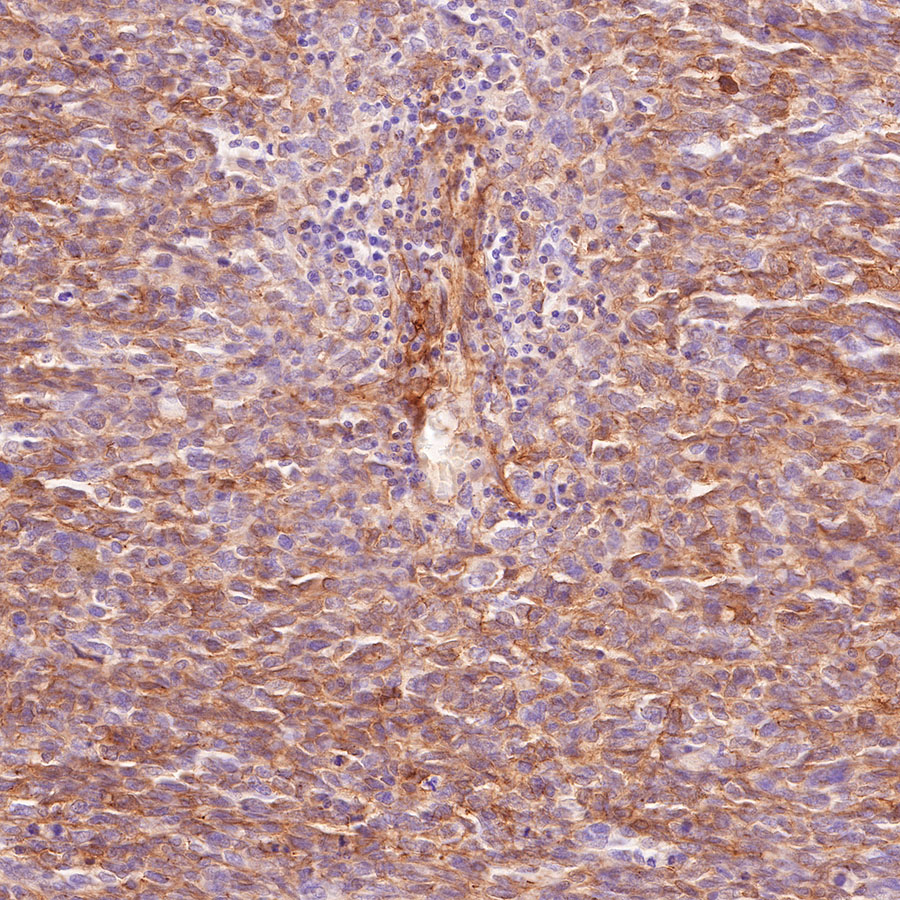
IHC shows positive staining in paraffin-embedded human melanoma. Anti-CD146 antibody was used at 1/1000 dilution, followed by a HRP Polymer for Mouse & Rabbit IgG (ready to use). Counterstained with hematoxylin. Heat mediated antigen retrieval with Tris/EDTA buffer pH9.0 was performed before commencing with IHC staining protocol.

IHC shows positive staining in paraffin-embedded human melanoma. Anti-CD146 antibody was used at 1/1000 dilution, followed by a HRP Polymer for Mouse & Rabbit IgG (ready to use). Counterstained with hematoxylin. Heat mediated antigen retrieval with Tris/EDTA buffer pH9.0 was performed before commencing with IHC staining protocol.

IHC shows positive staining in paraffin-embedded human breast cancer. Anti-CD146 antibody was used at 1/2000 dilution, followed by a HRP Polymer for Mouse & Rabbit IgG (ready to use). Counterstained with hematoxylin. Heat mediated antigen retrieval with Tris/EDTA buffer pH9.0 was performed before commencing with IHC staining protocol.
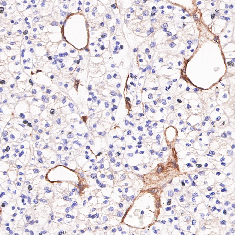
IHC shows positive staining in paraffin-embedded human renal clear cell carcinoma. Anti-CD146 antibody was used at 1/2000 dilution, followed by a HRP Polymer for Mouse & Rabbit IgG (ready to use). Counterstained with hematoxylin. Heat mediated antigen retrieval with Tris/EDTA buffer pH9.0 was performed before commencing with IHC staining protocol.

IHC shows positive staining in paraffin-embedded mouse lung. Anti-CD146 antibody was used at 1/2000 dilution, followed by a HRP Polymer for Mouse & Rabbit IgG (ready to use). Counterstained with hematoxylin. Heat mediated antigen retrieval with Tris/EDTA buffer pH9.0 was performed before commencing with IHC staining protocol.
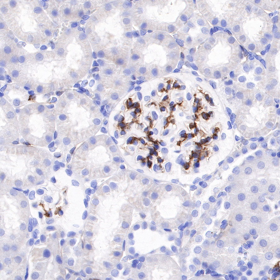
IHC shows positive staining in paraffin-embedded rat kidney. Anti-CD146 antibody was used at 1/2000 dilution, followed by a HRP Polymer for Mouse & Rabbit IgG (ready to use). Counterstained with hematoxylin. Heat mediated antigen retrieval with Tris/EDTA buffer pH9.0 was performed before commencing with IHC staining protocol.
 您现在的位置: