您现在的位置: 首页   -  产品中心   -  免疫学   -   抗体
S-RMab® CD16a Recombinant Rabbit mAb (SDT-366-45)

S-RMab® CD16a Recombinant Rabbit mAb (SDT-366-45)

货号: S0B2184
价格: 600
规格: 25μl
介绍: -
其他: -
产品规格
  • 宿主来源

    Rabbit
  • 抗原名称

    CD16a
  • 分子别名

    Low affinity immunoglobulin gamma Fc region receptor III-A, IgG Fc receptor III-A, CD16-II, Fc-gamma RIII-alpha, FcR-10, IgG Fc receptor III-2, FCGR3A
  • 免疫原

    Synthetic Peptide
  • 细胞定位

    Secreted, Cell membrane
  • Accession

    P08637
  • 克隆号

    SDT-366-45
  • 抗体类型

    Recombinant mAb
  • 应用

    ICFCM, IHC-P, IF
  • 反应种属 ?

    Hu, Ms, Rt
  • 纯化方式

    Protein A
  • 浓度

    0.5 mg/ml
  • 性状

    Liquid
  • 缓冲体系

    PBS, 40% Glycerol, 0.05% BSA, 0.03% Proclin 300
  • 储存条件

    12 months from date of receipt / reconstitution, -20 °C as supplied
稀释度
应用 稀释度
IHC-P 1:500
ICFCM 1:50
IF 1:200
背景介绍
  • CD16a is a transmembrane protein that acts as a receptor for the Fc portion of IgG antibodies. It is expressed on natural killer cells, monocytes, macrophages, dendritic cells, and some T cells. CD16a is also known as FCGR3A or FcγRIIIa. CD16a is involved in antibody dependent cellular cytotoxicity (ADCC) by binding to antibodies that target infected or malignant cells.

  • 流式分析

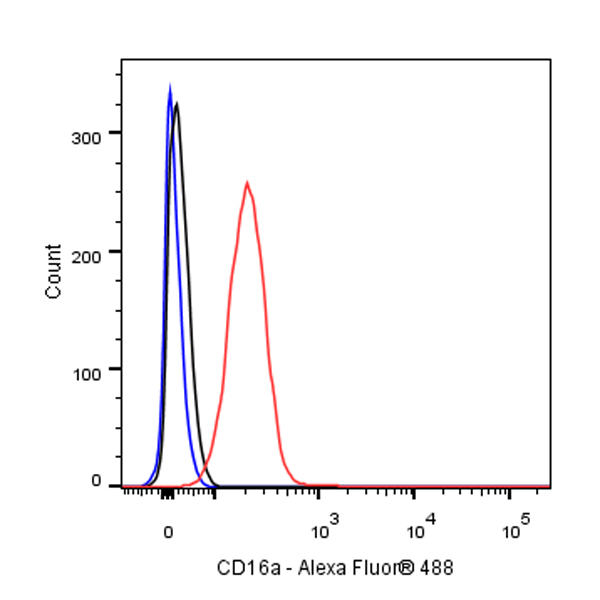
    • Flow cytometric analysis of 4% PFA fixed 90% methanol permeabilized Jurkat (Human T cell leukemia T lymphocyte) labelling CD16a antibody at 1/50 dilution (1 μg) / (Red) compared with a Rabbit monoclonal IgG (Black) isotype control and an unlabelled control (cells without incubation with primary antibody and secondary antibody) (Blue). Goat Anti - Rabbit IgG Alexa Fluor® 488 was used as the secondary antibody.

  • 免疫组化

    • IHC shows positive staining in paraffin-embedded human spleen. Anti-CD16a antibody was used at 1/500 dilution, followed by a HRP Polymer for Mouse & Rabbit IgG (ready to use). Counterstained with hematoxylin. Heat mediated antigen retrieval with Tris/EDTA buffer pH9.0 was performed before commencing with IHC staining protocol.
    • IHC shows positive staining in paraffin-embedded human liver. Anti-CD16a antibody was used at 1/500 dilution, followed by a HRP Polymer for Mouse & Rabbit IgG (ready to use). Counterstained with hematoxylin. Heat mediated antigen retrieval with Tris/EDTA buffer pH9.0 was performed before commencing with IHC staining protocol.
    • IHC shows positive staining in paraffin-embedded human lung. Anti-CD16a antibody was used at 1/500 dilution, followed by a HRP Polymer for Mouse & Rabbit IgG (ready to use). Counterstained with hematoxylin. Heat mediated antigen retrieval with Tris/EDTA buffer pH9.0 was performed before commencing with IHC staining protocol.
    • IHC shows positive staining in paraffin-embedded human lung cancer. Anti-CD16a antibody was used at 1/500 dilution, followed by a HRP Polymer for Mouse & Rabbit IgG (ready to use). Counterstained with hematoxylin. Heat mediated antigen retrieval with Tris/EDTA buffer pH9.0 was performed before commencing with IHC staining protocol.
    • IHC shows positive staining in paraffin-embedded human Hodgkin's lymphoma. Anti-CD16a antibody was used at 1/500 dilution, followed by a HRP Polymer for Mouse & Rabbit IgG (ready to use). Counterstained with hematoxylin. Heat mediated antigen retrieval with Tris/EDTA buffer pH9.0 was performed before commencing with IHC staining protocol.
    • IHC shows positive staining in paraffin-embedded human cervical squamous cell carcinoma. Anti-CD16a antibody was used at 1/500 dilution, followed by a HRP Polymer for Mouse & Rabbit IgG (ready to use). Counterstained with hematoxylin. Heat mediated antigen retrieval with Tris/EDTA buffer pH9.0 was performed before commencing with IHC staining protocol.
    • Negative control: IHC shows negative staining in paraffin-embedded human cardiac muscle. Anti-CD16a antibody was used at 1/500 dilution, followed by a HRP Polymer for Mouse & Rabbit IgG (ready to use). Counterstained with hematoxylin. Heat mediated antigen retrieval with Tris/EDTA buffer pH9.0 was performed before commencing with IHC staining protocol.
    • IHC shows positive staining in paraffin-embedded mouse liver. Anti-CD16a antibody was used at 1/500 dilution, followed by a HRP Polymer for Mouse & Rabbit IgG (ready to use). Counterstained with hematoxylin. Heat mediated antigen retrieval with Tris/EDTA buffer pH9.0 was performed before commencing with IHC staining protocol.
    • IHC shows positive staining in paraffin-embedded mouse stomach. Anti-CD16a antibody was used at 1/500 dilution, followed by a HRP Polymer for Mouse & Rabbit IgG (ready to use). Counterstained with hematoxylin. Heat mediated antigen retrieval with Tris/EDTA buffer pH9.0 was performed before commencing with IHC staining protocol.
    • IHC shows positive staining in paraffin-embedded rat liver. Anti-CD16a antibody was used at 1/500 dilution, followed by a HRP Polymer for Mouse & Rabbit IgG (ready to use). Counterstained with hematoxylin. Heat mediated antigen retrieval with Tris/EDTA buffer pH9.0 was performed before commencing with IHC staining protocol.
    • IHC shows positive staining in paraffin-embedded rat spleen. Anti-CD16a antibody was used at 1/500 dilution, followed by a HRP Polymer for Mouse & Rabbit IgG (ready to use). Counterstained with hematoxylin. Heat mediated antigen retrieval with Tris/EDTA buffer pH9.0 was performed before commencing with IHC staining protocol.
  • 免疫荧光

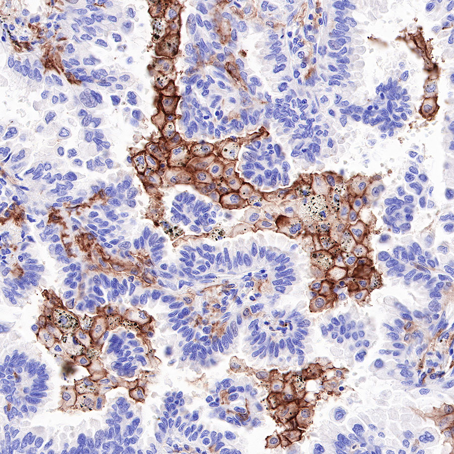
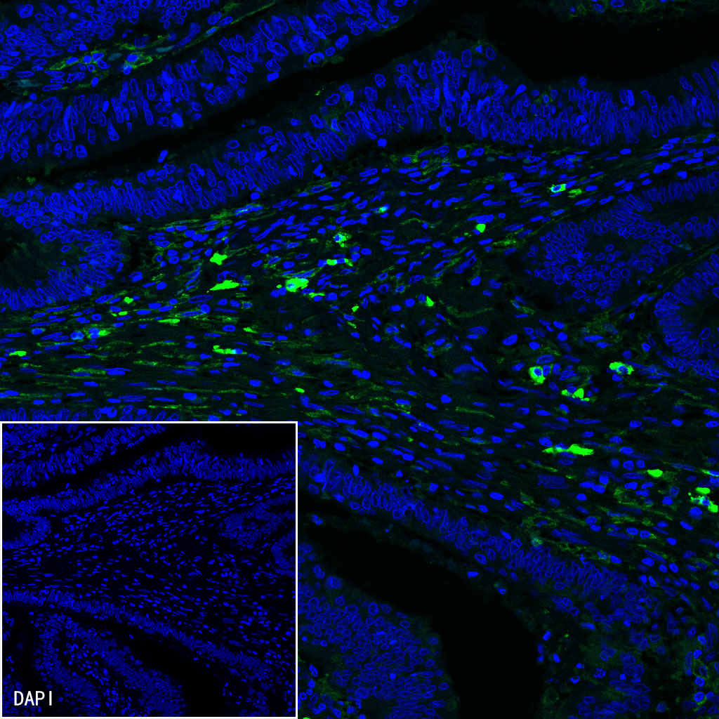
    • IF shows positive staining in paraffin-embedded human colon cancer. Anti-CD16a antibody was used at 1/200 dilution (Green) and incubated overnight at 4°C. Goat polyclonal Antibody to Rabbit IgG - H&L (Alexa Fluor® 488) was used as secondary antibody at 1/1000 dilution. Counterstained with DAPI (Blue). Heat mediated antigen retrieval with EDTA buffer pH9.0 was performed before commencing with IF staining protocol.

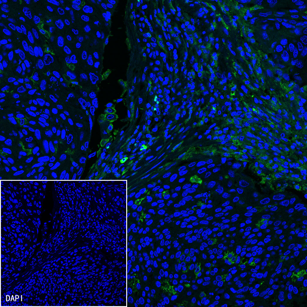
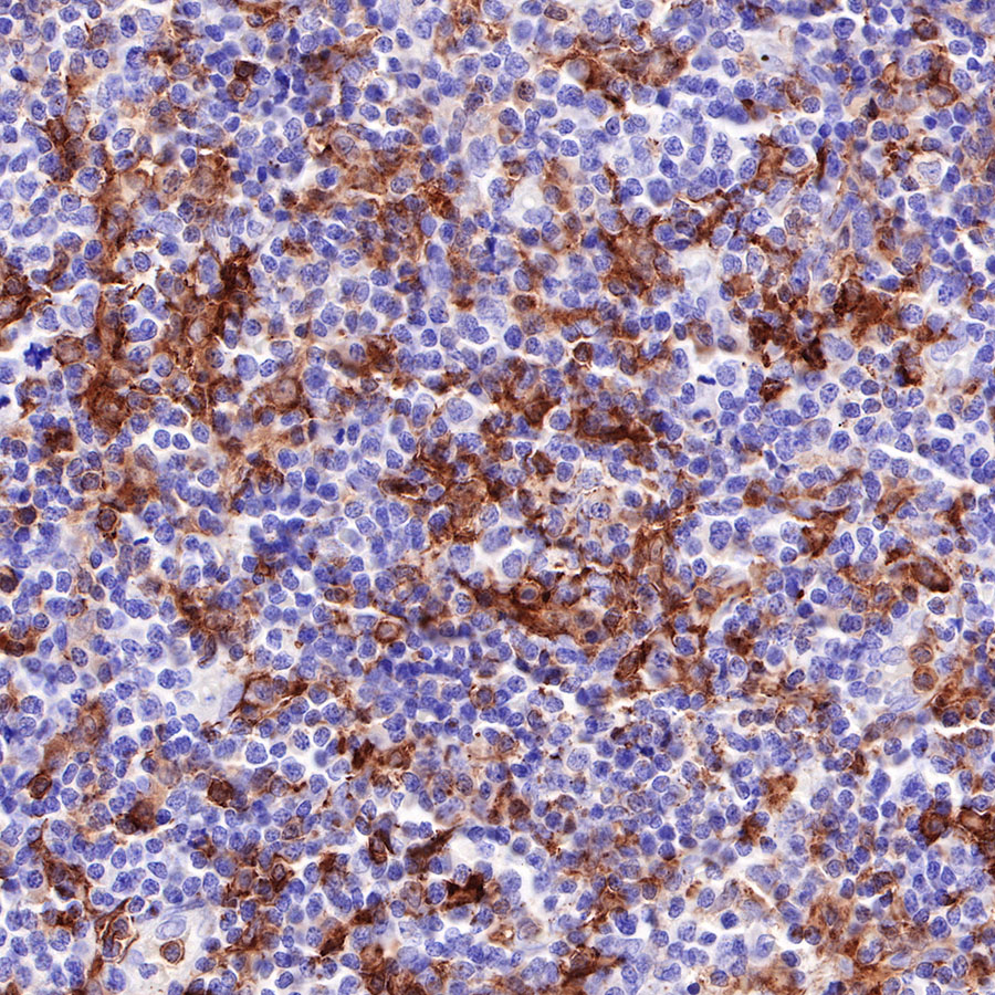
    • IF shows positive staining in paraffin-embedded human cervical squamous cell carcinoma. Anti-CD16a antibody was used at 1/200 dilution (Green) and incubated overnight at 4°C. Goat polyclonal Antibody to Rabbit IgG - H&L (Alexa Fluor® 488) was used as secondary antibody at 1/1000 dilution. Counterstained with DAPI (Blue). Heat mediated antigen retrieval with EDTA buffer pH9.0 was performed before commencing with IF staining protocol.

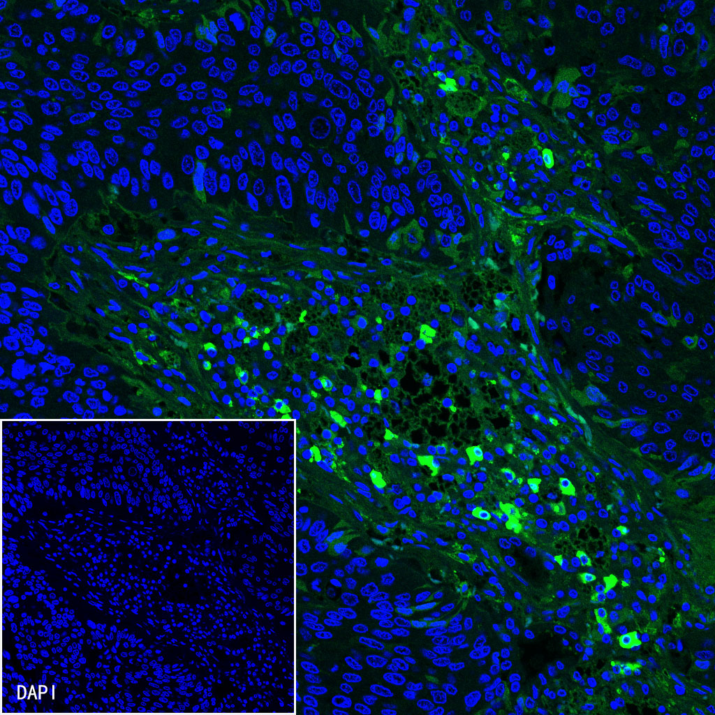
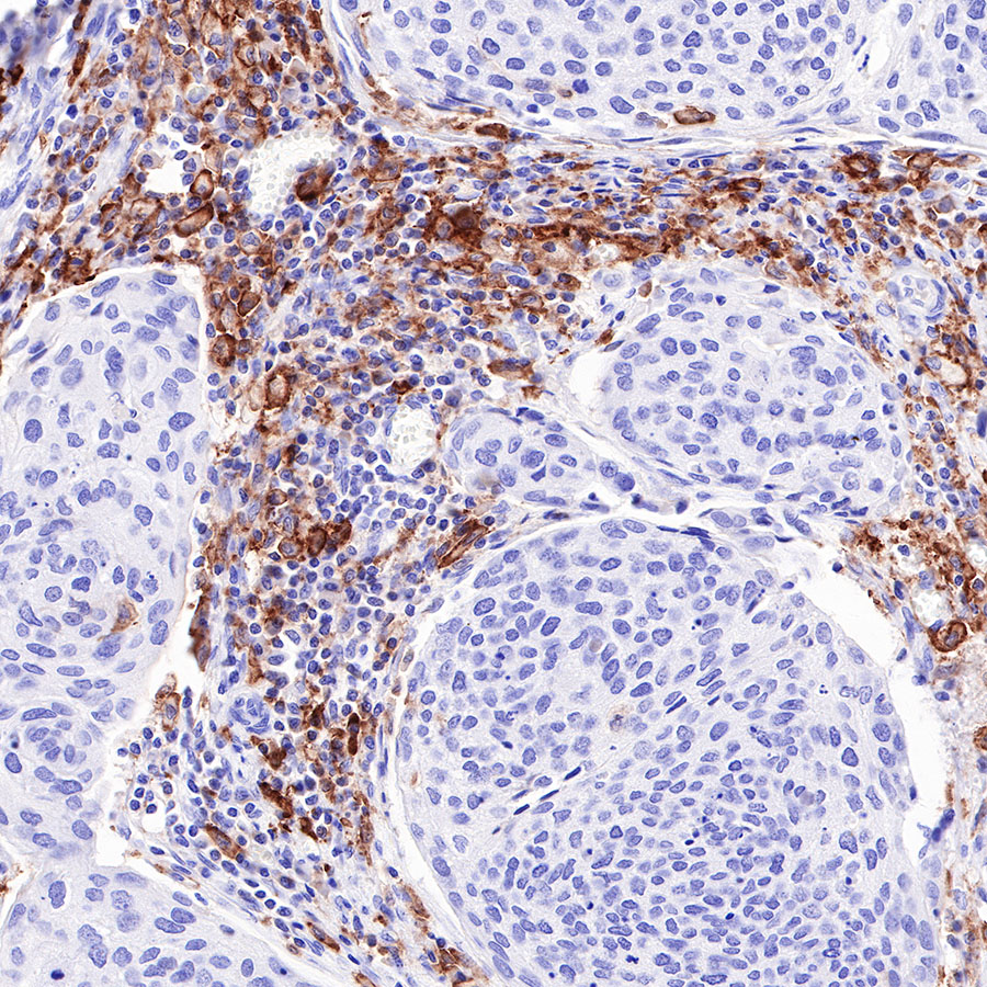
    • IF shows positive staining in paraffin-embedded human lung squamous cell carcinoma. Anti-CD16a antibody was used at 1/200 dilution (Green) and incubated overnight at 4°C. Goat polyclonal Antibody to Rabbit IgG - H&L (Alexa Fluor® 488) was used as secondary antibody at 1/1000 dilution. Counterstained with DAPI (Blue). Heat mediated antigen retrieval with EDTA buffer pH9.0 was performed before commencing with IF staining protocol.