
12 months from date of receipt / reconstitution, -20 °C as supplied
| 应用 | 稀释度 |
|---|---|
| WB | 1:1000 |
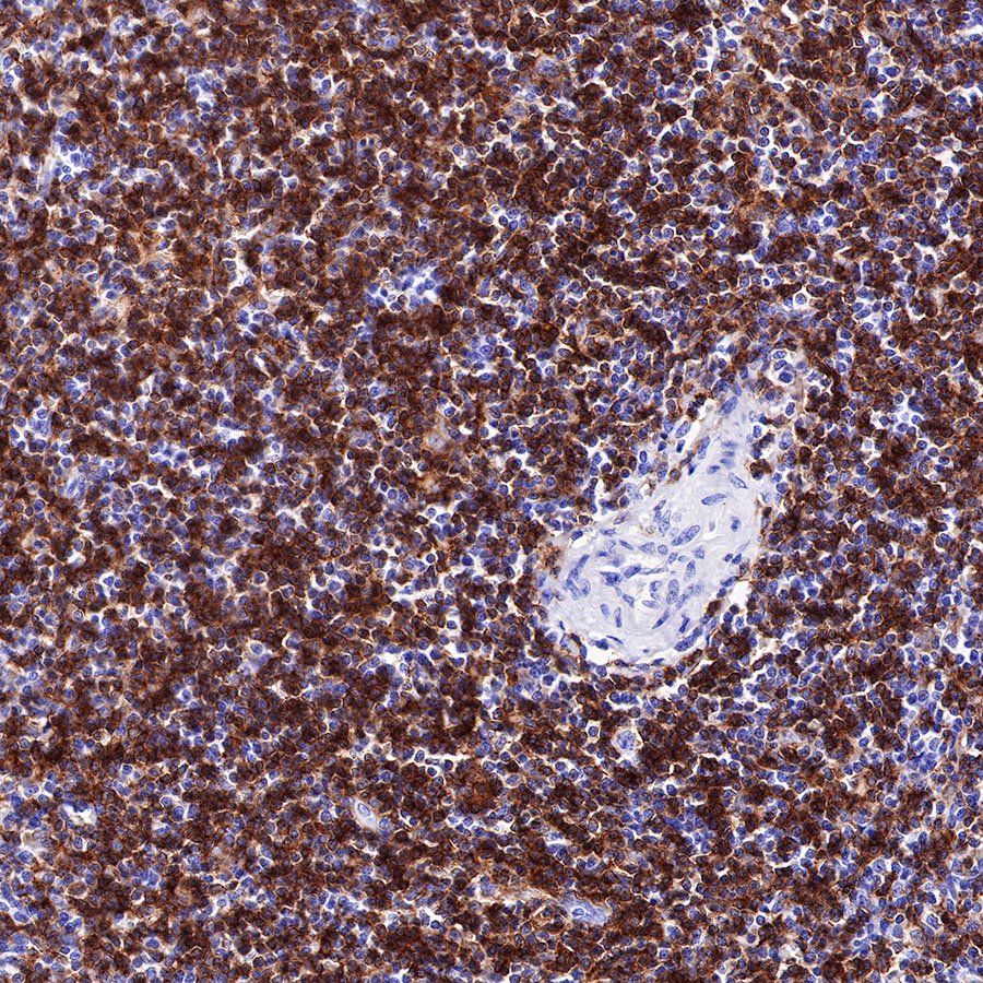
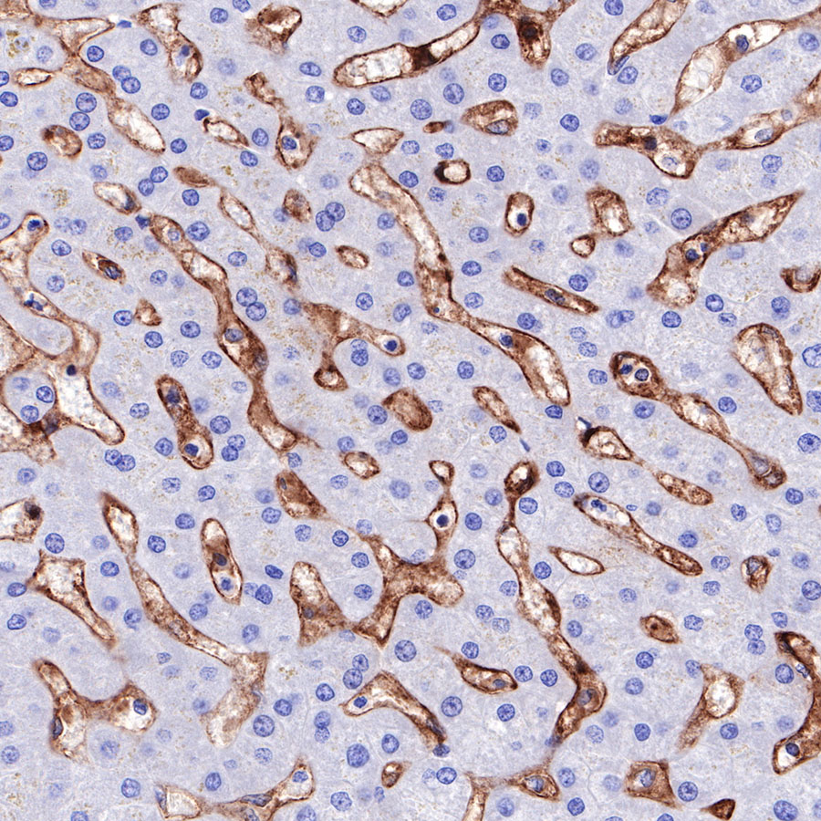
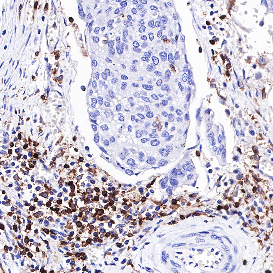
| IHC-P | 1:500 |
| FCM | 1:50 |
| IP | 1:50 |
| IF | 1:200 |
CD4 (cluster of differentiation 4) is a glycoprotein found on the surface of immune cells such as T helper cells, monocytes, macrophages, and dendritic cells. It is a type of white blood cell that helps fight infection by triggering your immune system to destroy viruses, bacteria, and other germs that may make you sick. CD4 is also a receptor for the HIV virus, and when the virus infects cells with CD4 surface proteins, it depletes the number of T cells, B cells, natural killer cells, and monocytes in the patient's blood.

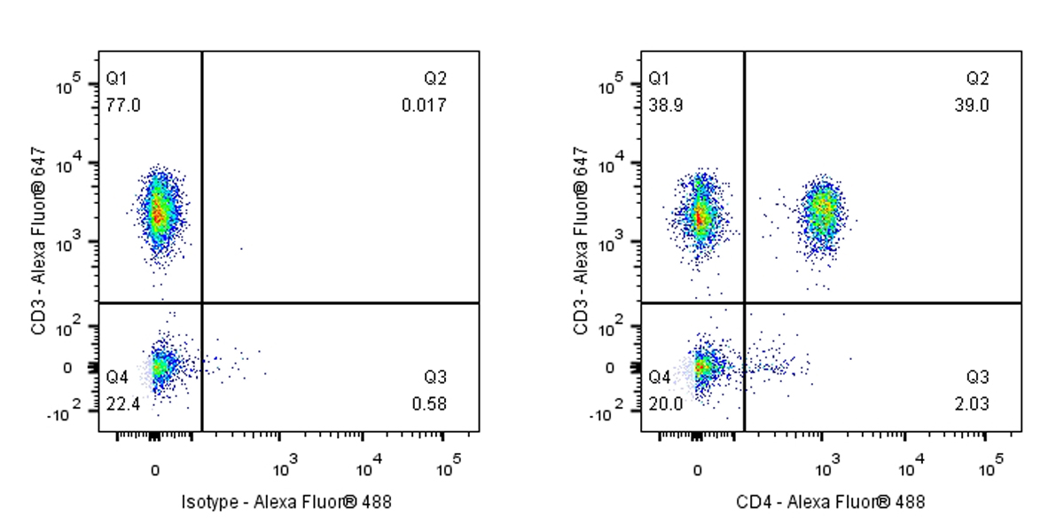
Flow cytometric analysis of human PBMC (human peripheral blood mononuclear cell) labelling CD4 antibody at 1/50 (1 μg) dilution (Right) compared with a Rabbit monoclonal IgG isotype control (Left). Goat Anti – Rabbit IgG Alexa Fluor® 488 was used as the secondary antibody, cells were stained with CD3 - Alexa Fluor® 647 simultaneously. Events were gated on viable lymphocytes.

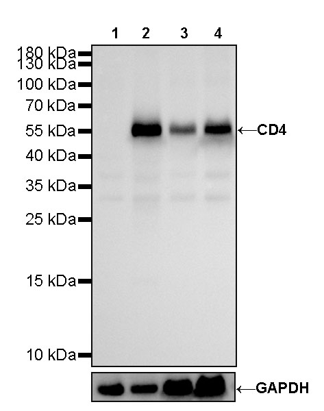
CD4 Rabbit mAb at 1/50 dilution (1 µg) immunoprecipitating CD4 in 0.4 mg THP-1 whole cell lysate.
Western blot was performed on the immunoprecipitate using CD4 Rabbit mAb at 1/1000 dilution.
Secondary antibody (HRP) for IP was used at 1/400 dilution.
Lane 1: THP-1 whole cell lysate 20 µg (Input)
Lane 2: CD4 Rabbit mAb IP in THP-1 whole cell lysate
Lane 3: Rabbit monoclonal IgG IP in THP-1 whole cell lysate
Predicted MW: 51 kDa
Observed MW: 55 kDa
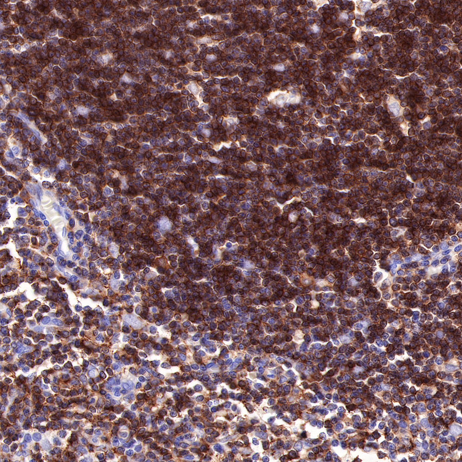
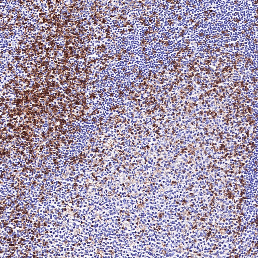

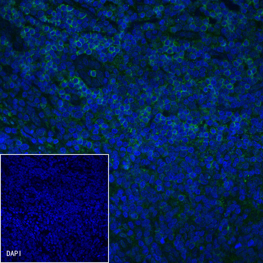
IF shows positive staining in paraffin-embedded human tonsil. Anti-CD4 antibody was used at 1/200 dilution (Green) and incubated overnight at 4°C. Goat polyclonal Antibody to Rabbit IgG - H&L (Alexa Fluor® 488) was used as secondary antibody at 1/1000 dilution. Counterstained with DAPI (Blue). Heat mediated antigen retrieval with EDTA buffer pH9.0 was performed before commencing with IF staining protocol.
 您现在的位置: