
12 months from date of receipt / reconstitution, -20 °C as supplied
| 应用 | 稀释度 |
|---|---|
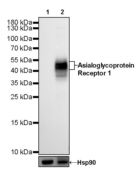
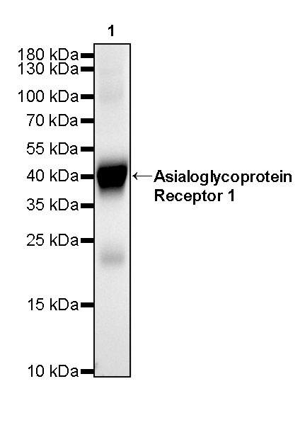
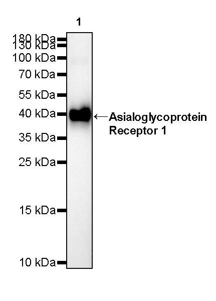
| WB | 1:1000-1:5000 |
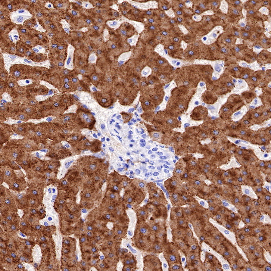
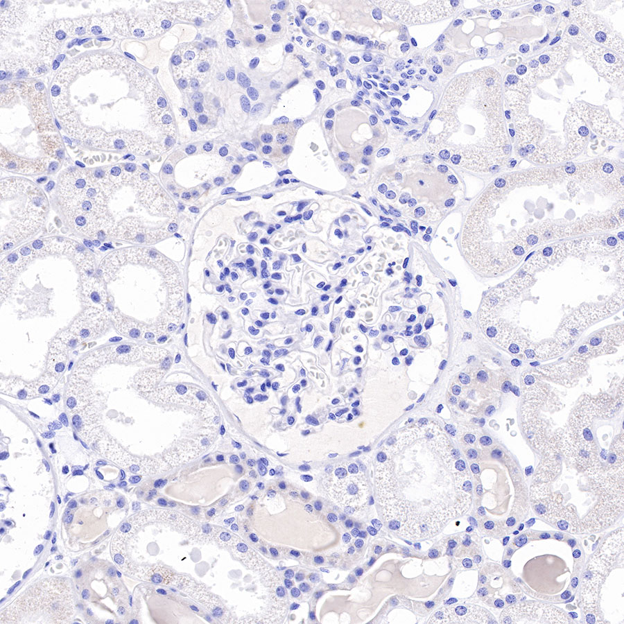
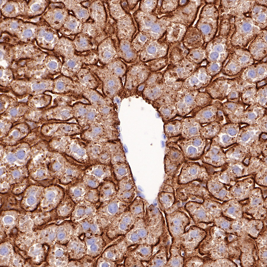
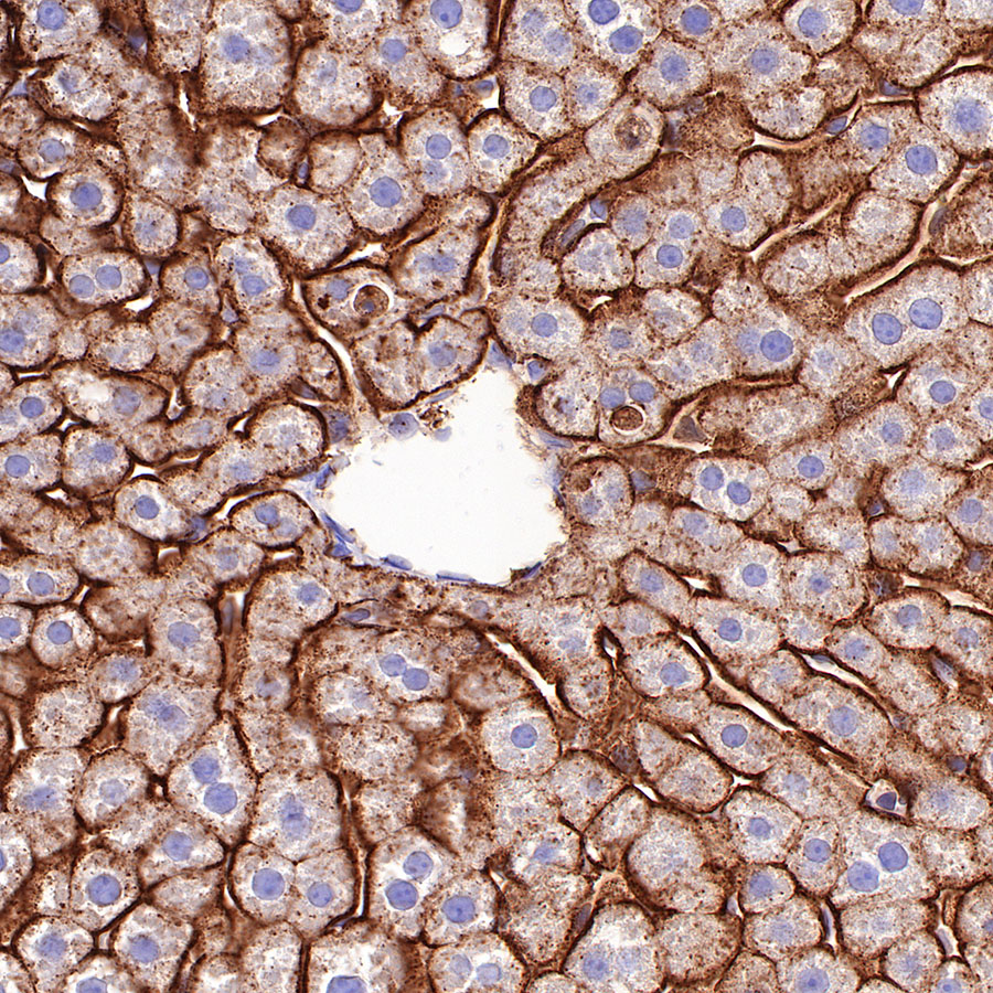
| IHC-P | 1:2000 |
| ICFCM | 1:5000 |
| ICC | 1:500 |
Asialoglycoprotein receptors (ASGP-R; exist as subtypes 1 and 2) are located on the surface of hepatic cell membranes and are involved in the binding and endocytosis of glycoproteins that have exposed carbohydrate or N-acetylgalactosamine residues. In addition, the decreased level of ASGP-R on the hepatic cells has been shown to correlate clinically with the degree of liver function retained due to the development of cirrhosis, cancer, or viral hepatitis [PMID: 20641215].


Flow cytometric analysis of 4% PFA fixed 90% methanol permeabilized HeLa (Human cervix adenocarcinoma epithelial cell, left) / HepG2 (Human hepatocellular carcinoma epithelial cell, Right) cells labelling Asialoglycoprotein Receptor 1 antibody at 1/5000 dilution (0.01 μg) / (Red) compared with a Rabbit monoclonal IgG (Black) isotype control and an unlabelled control (cells without incubation with primary antibody and secondary antibody) (Blue). Goat Anti - Rabbit IgG Alexa Fluor® 488 was used as the secondary antibody.Negative control: HeLa


ICC shows positive staining in HepG2 cells. Anti-ASGR1 antibody was used at 1/500 dilution (Green) and incubated overnight at 4°C. Goat polyclonal Antibody to Rabbit IgG - H&L (Alexa Fluor® 488) was used as secondary antibody at 1/1000 dilution. The cells were fixed with 100% ice-cold methanol and permeabilized with 0.1% PBS-Triton X-100. Nuclei were counterstained with DAPI (Blue).Counterstain with tubulin (Red).

Negative control:ICC shows negative staining in HeLa cells. Anti-ASGR1 antibody was used at 1/500 dilution and incubated overnight at 4°C. Goat polyclonal Antibody to Rabbit IgG - H&L (Alexa Fluor® 488) was used as secondary antibody at 1/1000 dilution. The cells were fixed with 100% ice-cold methanol and permeabilized with 0.1% PBS-Triton X-100. Nuclei were counterstained with DAPI (Blue).Counterstain with tubulin (Red).
 您现在的位置: