
PBS, 40% Glycerol, 0.05% BSA, 0.03% Proclin 300
12 months from date of receipt / reconstitution, -20 °C as supplied
| 应用 | 稀释度 |
|---|---|
| WB | 1:1000 |
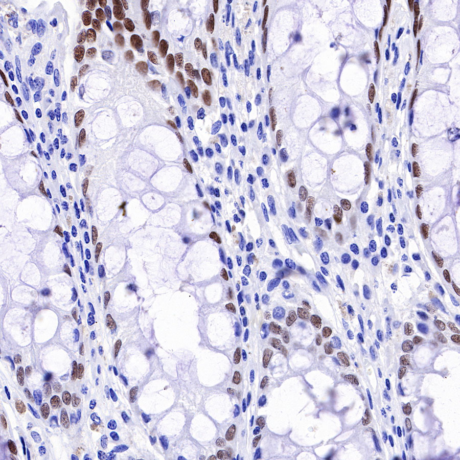
| IHC-P | 1:500 |
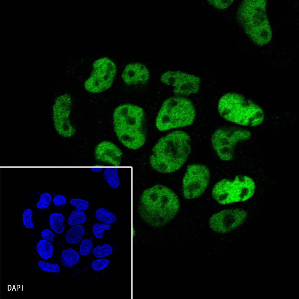
| ICC | 1:500 |
| IP | 1:50 |
| ICFCM | 1:500 |
HNF4α is a transcription factor that plays an important role in liver morphogenesis and maintenance of proper hepatocyte function in a mature liver. Most liver diseases have been associated with altered HNF4α expression, isoform ratios, and localization. Therefore, many studies have indicated that HNF4α may potentially be a target gene for the regression of fibrosis and cirrhosis [PMID: 35114889].


Flow cytometric analysis of 4% PFA fixed 90% methanol permeabilized HeLa (Left) / HepG2 (Right) cells labelling HNF4α antibody at 1/500 dilution (0.1 μg)/ (Red) compared with a Rabbit monoclonal IgG (Black) isotype control and an unlabelled control (cells without incubation with primary antibody and secondary antibody) (Blue). Goat Anti-Rabbit IgG Alexa Fluor® 488 was used as the secondary antibody. Negative control: HeLa
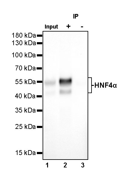
HNF4α Rabbit mAb at 1/50 dilution (1 µg) immunoprecipitating HNF4α in 0.4 mg HepG2 whole cell lysate.
Western blot was performed on the immunoprecipitate using HNF4α Rabbit mAb at 1/1000 dilution.
Secondary antibody (HRP) for IP was used at 1/400 dilution.
Lane 1: HepG2 whole cell lysate 20 µg (Input)
Lane 2: HNF4α Rabbit mAb IP in HepG2 whole cell lysate
Lane 3: Rabbit monoclonal IgG IP in HepG2 whole cell lysate
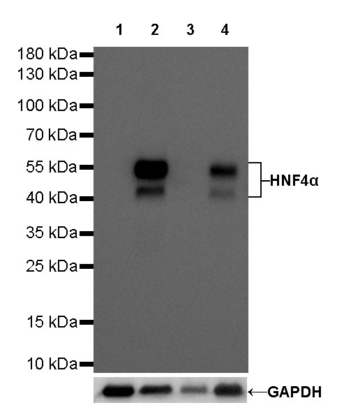
Predicted MW: 53 kDa
Observed MW: 45~55 kDa
Exposure time: 20 s


 您现在的位置: