

PBS, 40% Glycerol, 0.05% BSA, 0.03% Proclin 300
12 months from date of receipt / reconstitution, -20 °C as supplied
| 应用 | 稀释度 |
|---|---|
| WB | 1:1000 |
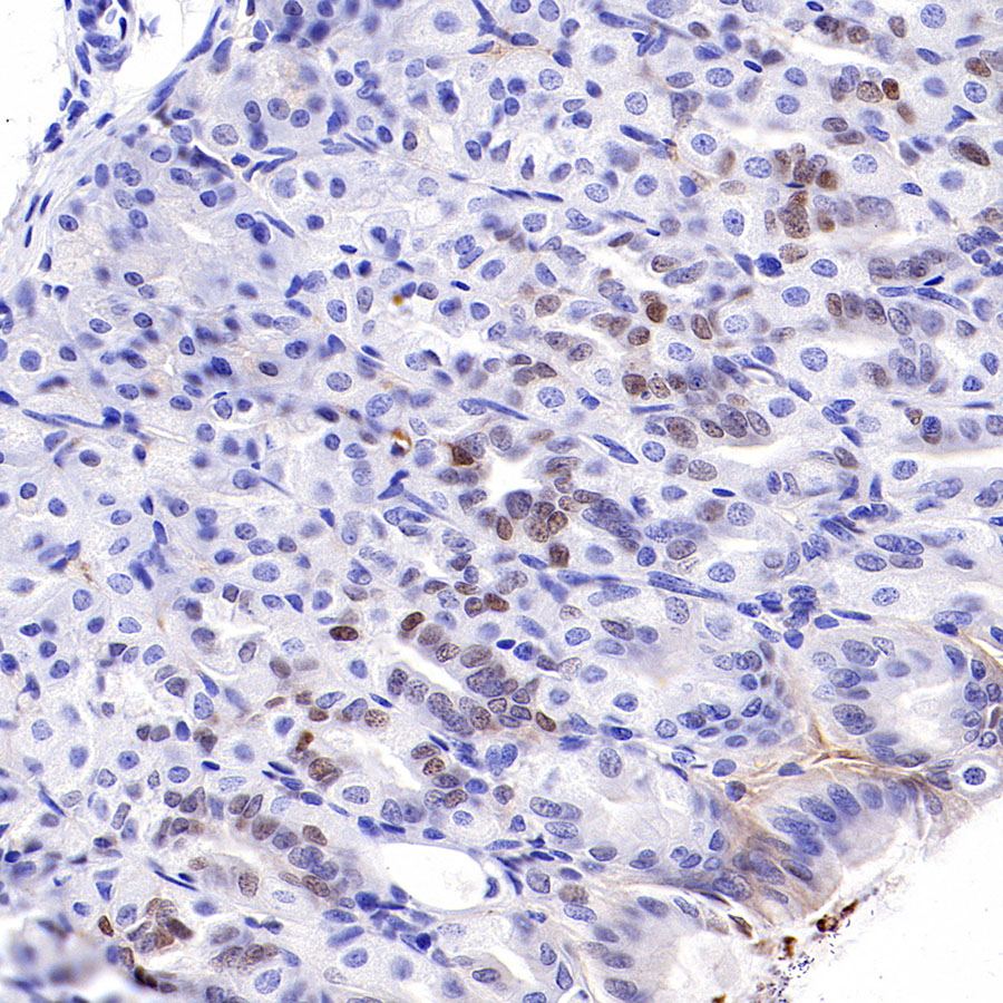
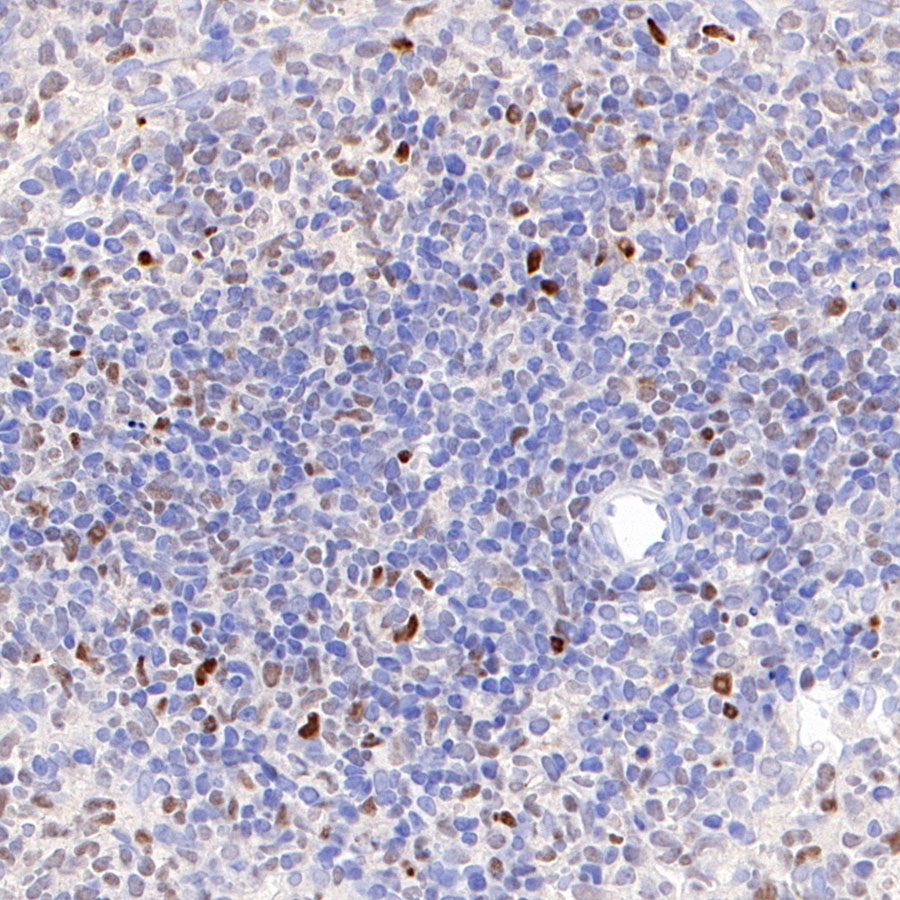
| IHC-P | 1:500-1:1000 |
| ICFCM | 1:50 |
| ICC | 1:500 |
c-Myc (MYC) is a helix-loop-helix leucine zipper transcription factor that dimerizes with its partner protein Max to bind specific DNA sequences and transactivates genes. The Myc-Max heterodimer can also repress gene expression through complex formation with the transcription factor Miz1 [PubMed: 18923074]. In addition to its role in cancer, Myc is one of four transcription factors that collectively can re-program differentiated adult cells back to a pluripotent stem cell state [PubMed: 16904174]. Myc also plays an important role in normal cell physiology. The key distinction between physiological and oncogenic Myc function is whether MYC expression is regulated by normal circuitries, such as growth factor signaling that occurs when cells enter into the cell cycle and proliferate for tissue repair or whether, as in cancers, MYC activation can be short-circuited by genetic alterations, permitting deregulated Myc expression to alter transcription that no longer responds to external cues, particularly negative regulatory ones [PubMed: 16267388].
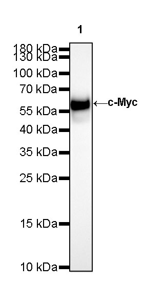
WB result of c-Myc Rabbit mAb
Primary antibody: c-Myc Rabbit mAb at 1/1000 dilution
Lane 1: Neuro-2a whole cell lysate 20 µg
Secondary antibody: Goat Anti-Rabbit IgG, (H+L), HRP conjugated at 1/10000 dilution
Predicted MW: 49 kDa
Observed MW: 60 kDa
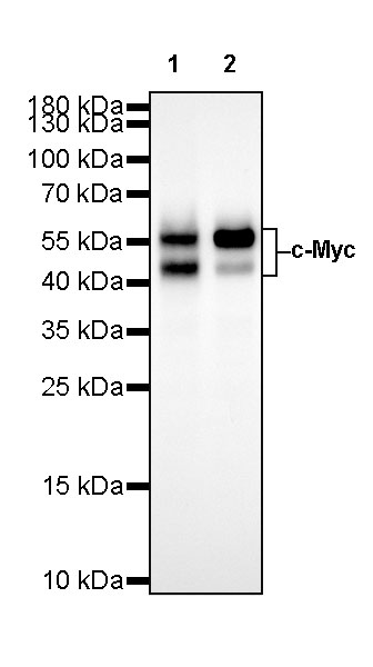
WB result of c-Myc Rabbit mAb
Primary antibody: c-Myc Rabbit mAb at 1/1000 dilution
Lane 1: HeLa whole cell lysate 20 µg
Lane 2: Jurkat whole cell lysate 20 µg
Secondary antibody: Goat Anti-Rabbit IgG, (H+L), HRP conjugated at 1/10000 dilution
Predicted MW: 49 kDa
Observed MW: 45, 57 kDa
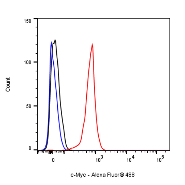
Flow cytometric analysis of 4% PFA fixed 90% methanol permeabilized HeLa (Human cervix adenocarcinoma epithelial cell) labelling c-Myc antibody at 1/50 dilution (1 μg)/ (Right) compared with a Rabbit monoclonal IgG / (Left) isotype control. Goat Anti - Rabbit IgG Alexa Fluor® 488 was used as the secondary antibody.

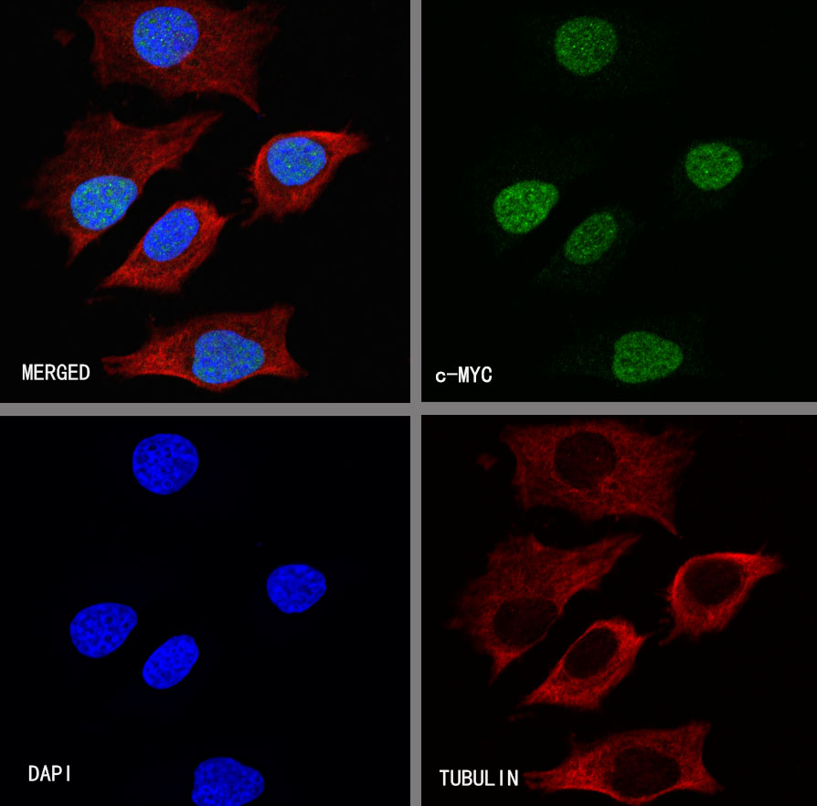
ICC shows positive staining in HeLa cells. Anti-c-Myc antibody was used at 1/500 dilution (Green) and incubated overnight at 4°C. Goat polyclonal Antibody to Rabbit IgG - H&L (Alexa Fluor® 488) was used as secondary antibody at 1/1000 dilution. The cells were fixed with 100% ice-cold methanol and permeabilized with 0.1% PBS-Triton X-100. Nuclei were counterstained with DAPI (Blue).Counterstain with tubulin (red).
 您现在的位置: