
PBS, 40% Glycerol, 0.05% BSA, 0.03% Proclin 300
12 months from date of receipt / reconstitution, -20 °C as supplied
| 应用 | 稀释度 |
|---|---|
| IHC-P | 1:1000 |
| WB | 1:500 |
| IP | 1:25 |
| ICC | 1:250 |
| IF | 1:500 |
The p53 protein is a crucial tumor suppressor encoded by the TP53 gene, named for its molecular weight of approximately 53 kilodaltons. Known as the "guardian of the genome," p53 is activated in response to cellular stress (such as DNA damage, hypoxia, or oncogene activation) and regulates the transcription of downstream target genes. This leads to cell cycle arrest, DNA repair, apoptosis, or senescence, thereby maintaining genomic stability and preventing tumorigenesis. Loss of p53 function (e.g., mutation or degradation) is associated with over 50% of human cancers, and mutant p53 may also acquire oncogenic properties. Its activity is primarily negatively regulated by E3 ubiquitin ligases like MDM2, which maintain low p53 levels via ubiquitin-mediated degradation. Beyond tumor suppression, p53 is involved in metabolic regulation, immune responses, and other processes, making it a key therapeutic target in cancer. Current p53-targeted strategies include restoring mutant p53 function, inhibiting MDM2, and exploiting p53-dependent pathways.
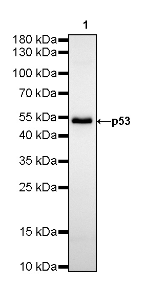
WB result of P53 Rabbit mAb
Primary antibody: P53 Rabbit mAb at 1/500 dilution
Lane 1: T-47D whole cell lysate 20 µg
Secondary antibody: Goat Anti-Rabbit IgG, (H+L), HRP conjugated at 1/10000 dilution
Predicted MW: 53 kDa
Observed MW: 53 kDa
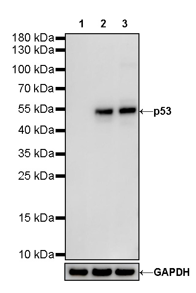
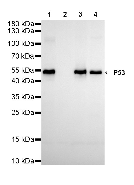
WB result of P53 Rabbit mAb
Primary antibody: P53 Rabbit mAb at 1/500 dilution
Lane 1: HL-60 whole cell lysate 20 µg
Lane 2: HT-29 whole cell lysate 20 µg
Lane 3: A431 whole cell lysate 20 µg
Negative control: HL-60 whole cell lysate
Secondary antibody: Goat Anti-Rabbit IgG, (H+L), HRP conjugated at 1/10000 dilution
Predicted MW: 53 kDa
Observed MW: 53 kDa
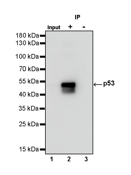
p53 Rabbit mAb at 1/25 dilution (1 µg) immunoprecipitating p53 in 0.4 mg A431 whole cell lysate.
Western blot was performed on the immunoprecipitate using p53 Rabbit mAb at 1/1000 dilution.
Secondary antibody (HRP) for IP was used at 1/400 dilution.
Lane 1: A431 whole cell lysate 20 µg (Input)
Lane 2: p53 Rabbit mAb IP in A431 whole cell lysate
Lane 3: Rabbit monoclonal IgG IP in A431 whole cell lysate
Predicted MW: 53 kDa
Observed MW: 53 kDa
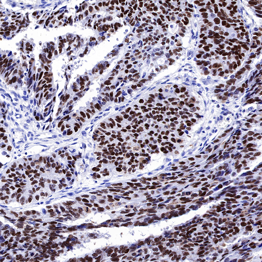
IHC shows positive staining in paraffin-embedded human colon cancer. Anti-p53 antibody was used at 1/1000 dilution, followed by a HRP Polymer for Mouse & Rabbit IgG (ready to use). Counterstained with hematoxylin. Heat mediated antigen retrieval with Tris/EDTA buffer pH9.0 was performed before commencing with IHC staining protocol.
IHC shows positive staining in paraffin-embedded human cervical squamous cell carcinoma. Anti-p53 antibody was used at 1/1000 dilution, followed by a HRP Polymer for Mouse & Rabbit IgG (ready to use). Counterstained with hematoxylin. Heat mediated antigen retrieval with Tris/EDTA buffer pH9.0 was performed before commencing with IHC staining protocol.
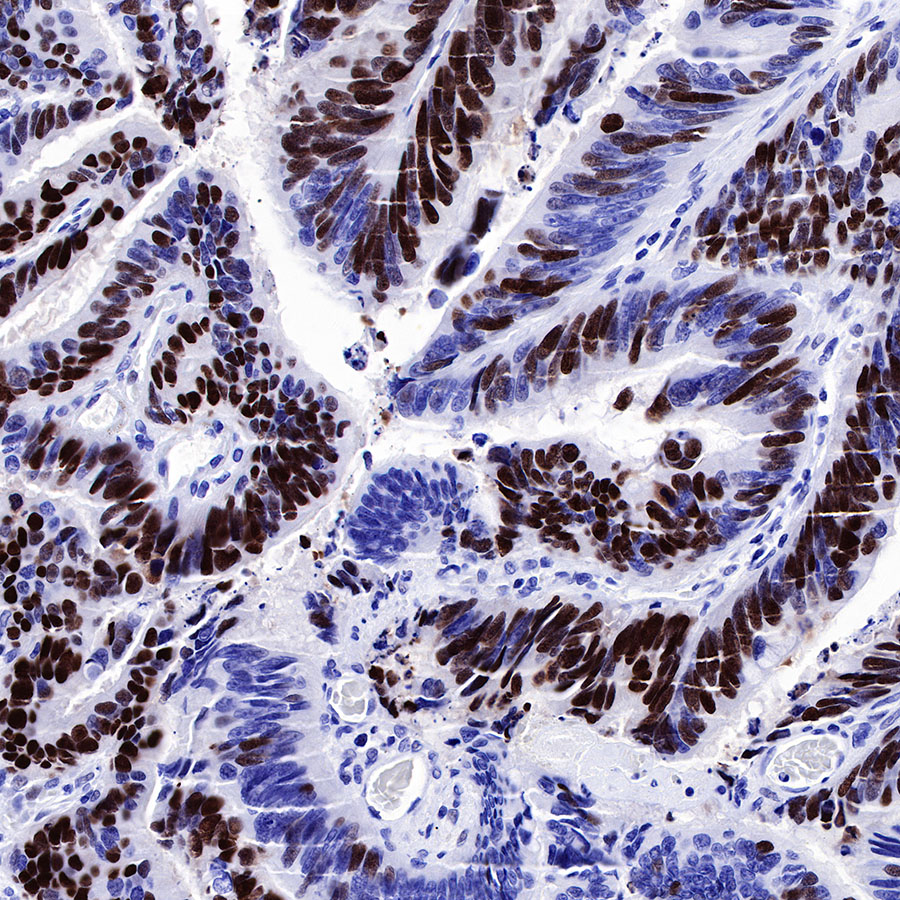
IHC shows positive staining in paraffin-embedded human colon cancer. Anti-p53 antibody was used at 1/1000 dilution, followed by a HRP Polymer for Mouse & Rabbit IgG (ready to use). Counterstained with hematoxylin. Heat mediated antigen retrieval with Tris/EDTA buffer pH9.0 was performed before commencing with IHC staining protocol.
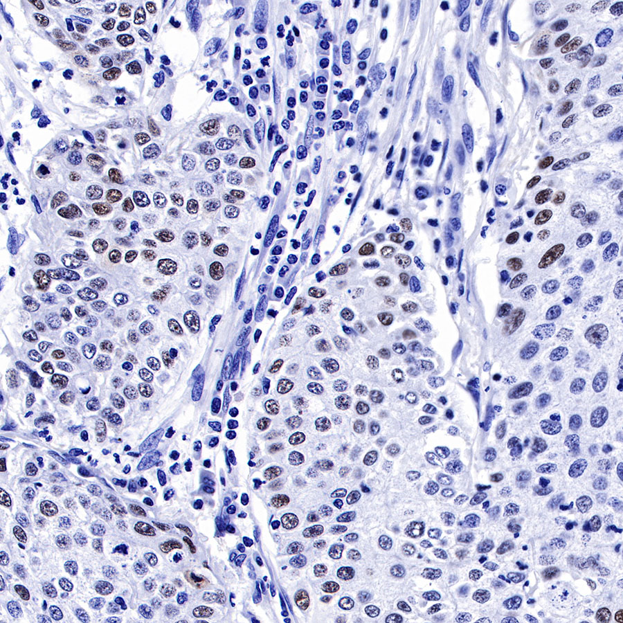
IHC shows positive staining in paraffin-embedded human lung squamous cell carcinoma. Anti-p53 antibody was used at 1/1000 dilution, followed by a HRP Polymer for Mouse & Rabbit IgG (ready to use). Counterstained with hematoxylin. Heat mediated antigen retrieval with Tris/EDTA buffer pH9.0 was performed before commencing with IHC staining protocol.
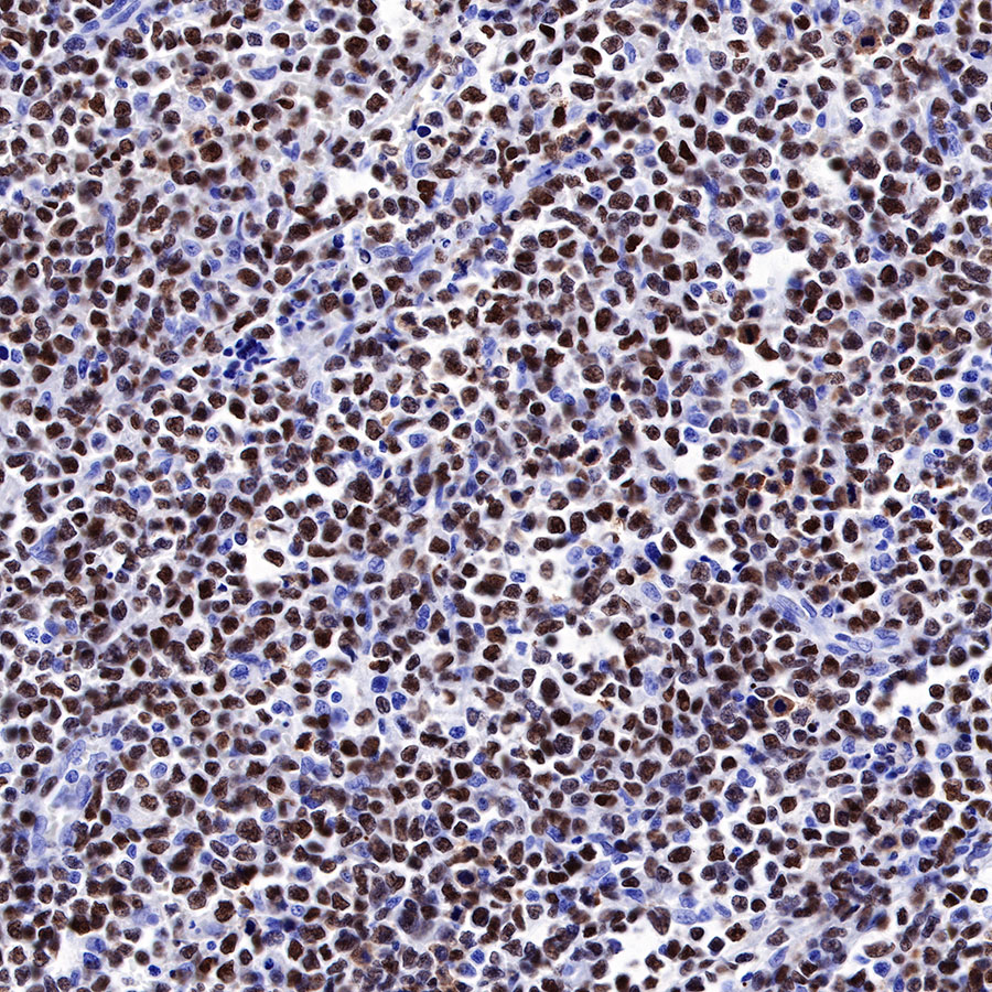
IHC shows positive staining in paraffin-embedded human NK/T-cell lymphoma. Anti-p53 antibody was used at 1/1000 dilution, followed by a HRP Polymer for Mouse & Rabbit IgG (ready to use). Counterstained with hematoxylin. Heat mediated antigen retrieval with Tris/EDTA buffer pH9.0 was performed before commencing with IHC staining protocol.
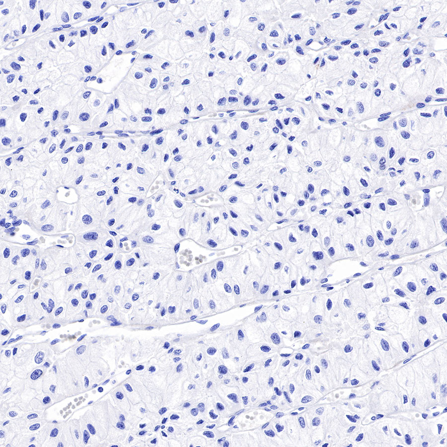
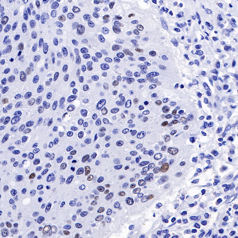
Negative control: IHC shows negative staining in paraffin-embedded human chromophobe renal cell carcinoma. Anti-p53 antibody was used at 1/1000 dilution, followed by a HRP Polymer for Mouse & Rabbit IgG (ready to use). Counterstained with hematoxylin. Heat mediated antigen retrieval with Tris/EDTA buffer pH9.0 was performed before commencing with IHC staining protocol.
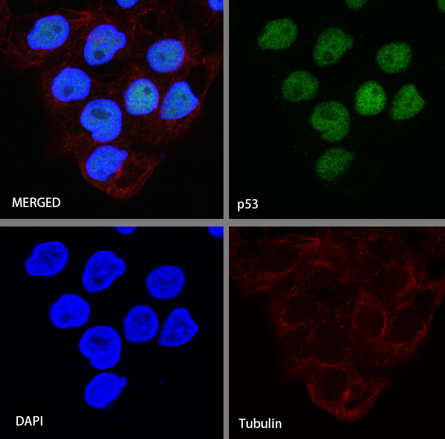
ICC shows positive staining in A431 cells. Anti-p53 antibody was used at 1/250 dilution (Green) and incubated overnight at 4°C. Goat polyclonal Antibody to Rabbit IgG - H&L (Alexa Fluor® 488) was used as secondary antibody at 1/1000 dilution. The cells were fixed with 4% PFA and permeabilized with 0.1% PBS-Triton X-100. Nuclei were counterstained with DAPI (Blue). Counterstain with tubulin (red).
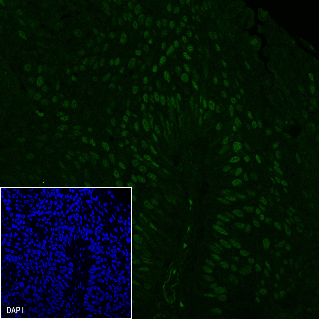
IF shows positive staining in paraffin-embedded human lung squamous cell carcinoma. Anti-p53 antibody was used at 1/500 dilution (Green) and incubated overnight at 4°C. Goat polyclonal Antibody to Rabbit IgG - H&L (Alexa Fluor® 488) was used as secondary antibody at 1/1000 dilution. Counterstained with DAPI (Blue). Heat mediated antigen retrieval with EDTA buffer pH9.0 was performed before commencing with IF staining protocol.
 您现在的位置: