
| 应用 | 稀释度 |
|---|---|
| IHC-P | 1:1000 |
| WB | 1:500 |
| ICC | 1:250 |
Calcitonin is a hormone that is produced in humans by the parafollicular cells (commonly known as C-cells) of the thyroid gland. It is found in the thyroid of chordates and humans and the endostyle of invertebrates. Calcitonin is involved in helping to regulate levels of calcium and phosphate in the blood, opposing the action of parathyroid hormone.

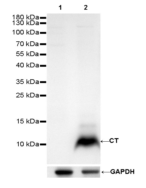
WB result of Calcitonin (CT) Rabbit mAb
Primary antibody: Calcitonin (CT) Rabbit mAb at 1/500 dilution
Lane 1: HeLa whole cell lysate 20 µg
Lane 2: TT whole cell lysate 20 µg
Negative control: HeLa whole cell lysate
Secondary antibody: Goat Anti-Rabbit IgG, (H+L), HRP conjugated at 1/10000 dilution
Predicted MW: 3.6 kDa
Observed MW: 10 kDa
Exposure time: 60s
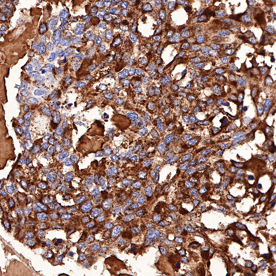
IHC shows positive staining in paraffin-embedded human medullary thyroid carcinoma. Anti-Calcitonin antibody was used at 1/1000 dilution, followed by a HRP Polymer for Mouse & Rabbit IgG (ready to use). Counterstained with hematoxylin. Heat mediated antigen retrieval with Tris/EDTA buffer pH9.0 was performed before commencing with IHC staining protocol.
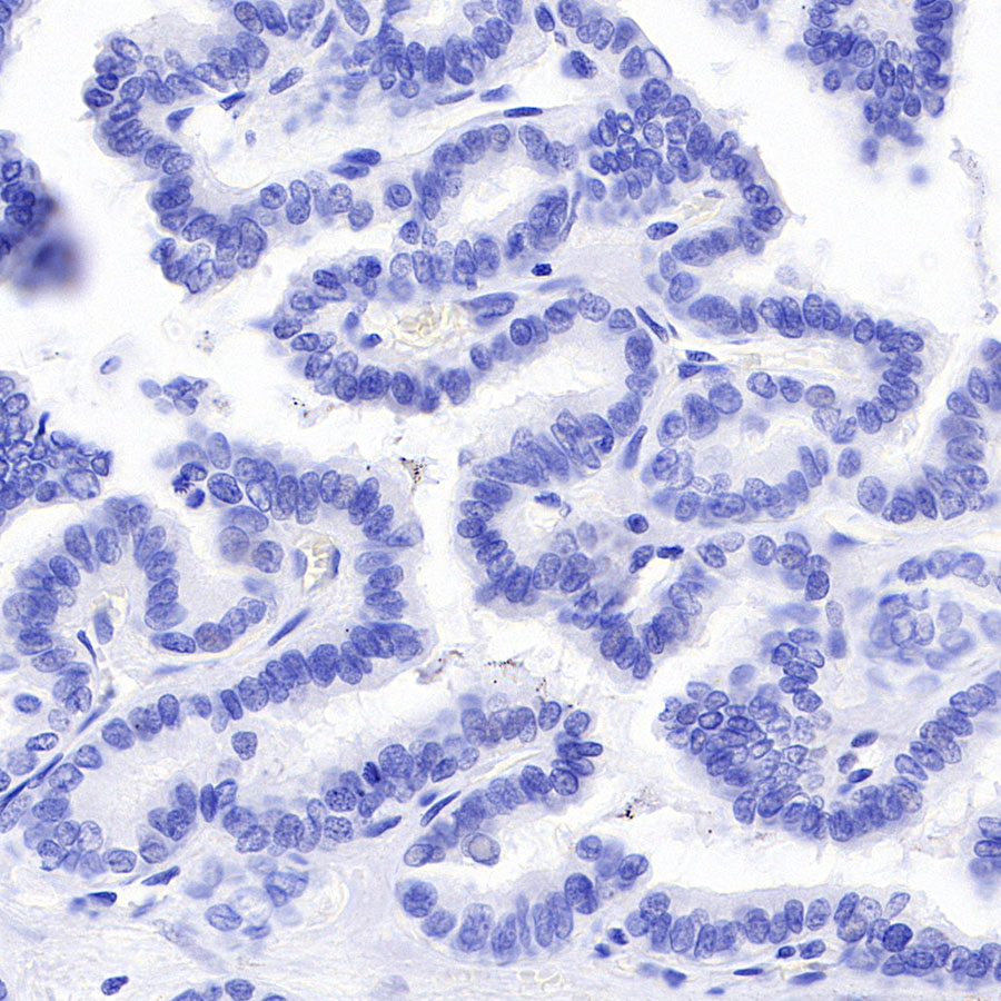
Negative control: IHC shows negative staining in paraffin-embedded human papillary thyroid carcinoma. Anti-Calcitonin antibody was used at 1/1000 dilution, followed by a HRP Polymer for Mouse & Rabbit IgG (ready to use). Counterstained with hematoxylin. Heat mediated antigen retrieval with Tris/EDTA buffer pH9.0 was performed before commencing with IHC staining protocol.
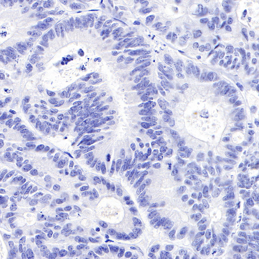
Negative control: IHC shows negative staining in paraffin-embedded human colon cancer. Anti-Calcitonin antibody was used at 1/1000 dilution, followed by a HRP Polymer for Mouse & Rabbit IgG (ready to use). Counterstained with hematoxylin. Heat mediated antigen retrieval with Tris/EDTA buffer pH9.0 was performed before commencing with IHC staining protocol.
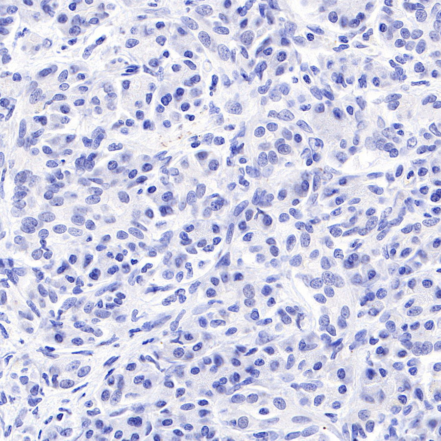
Negative control: IHC shows negative staining in paraffin-embedded human pancreatic cancer. Anti-Calcitonin antibody was used at 1/1000 dilution, followed by a HRP Polymer for Mouse & Rabbit IgG (ready to use). Counterstained with hematoxylin. Heat mediated antigen retrieval with Tris/EDTA buffer pH9.0 was performed before commencing with IHC staining protocol.
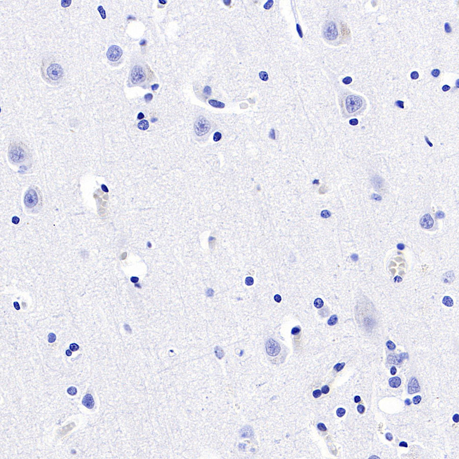
Negative control: IHC shows negative staining in paraffin-embedded human cerebral cortex. Anti-Calcitonin antibody was used at 1/1000 dilution, followed by a HRP Polymer for Mouse & Rabbit IgG (ready to use). Counterstained with hematoxylin. Heat mediated antigen retrieval with Tris/EDTA buffer pH9.0 was performed before commencing with IHC staining protocol.
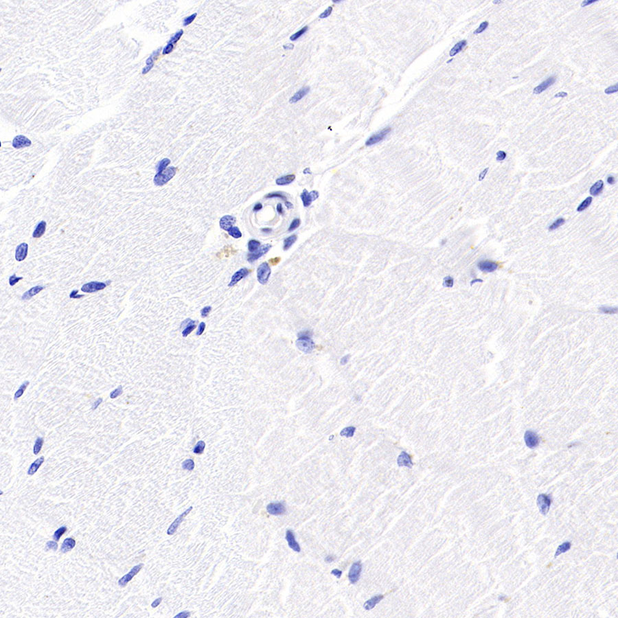
Negative control: IHC shows negative staining in paraffin-embedded human skeletal muscle. Anti-Calcitonin antibody was used at 1/1000 dilution, followed by a HRP Polymer for Mouse & Rabbit IgG (ready to use). Counterstained with hematoxylin. Heat mediated antigen retrieval with Tris/EDTA buffer pH9.0 was performed before commencing with IHC staining protocol.
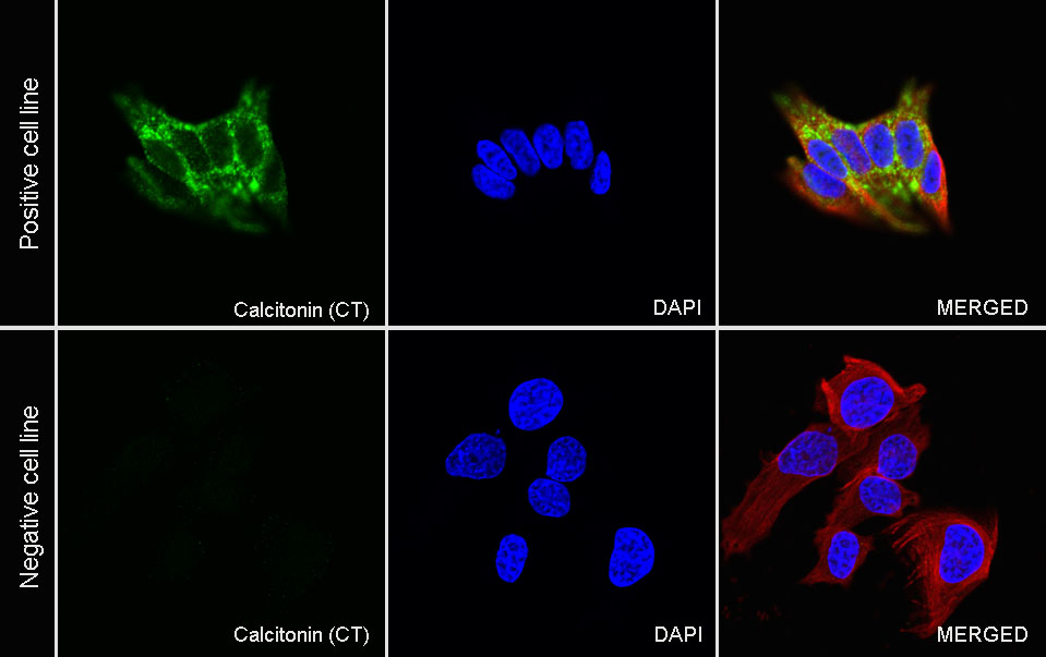
ICC shows positive staining in TT cells (top panel) and negative staining in HeLa cells (below panel). Anti-Calcitonin (CT) antibody was used at 1/250 dilution (Green) and incubated overnight at 4°C. Goat polyclonal Antibody to Rabbit IgG - H&L (Alexa Fluor® 488) was used as secondary antibody at 1/1000 dilution. The cells were fixed with 4% PFA and permeabilized with 0.1% PBS-Triton X-100. Nuclei were counterstained with DAPI (Blue). Counterstain with tubulin (Red).
 您现在的位置: