
12 months from date of receipt / reconstitution, -20 °C as supplied
| 应用 | 稀释度 |
|---|---|
| WB | 1:500-1:10000 |
| IHC-P | 1:1000 |
Caveolin-1 is a 22 kDa protein encoded by CAV1 gene, and occupies flask-shaped plasma membrane invaginations called caveolae. It is one of three known caveolins (CAV1, 2 and 3) and is ubiquitously expressed in all cell types as is CAV2; CAV3 is mostly found in skeletal muscles. Besides earlier studies that implicated CAV1 in endocytosis, signaling and lipid disorders, research activities in the last two decades also focused on clarifying its relevance in cancer. Caveolin-1 (Cav-1) has been considered as a master regulator among the various signaling molecules. It has been emerging as a chief protein regulating cellular events associated with homeostasis, caveolae formation, and caveolae trafficking. In addition to the physiological role of cav-1, it has a complex role in the progression of various diseases. Caveolin-1 has been identified as a prognosticator in patients with cancer and has a dual role in tumorigenesis. The expression of Cav-1 in hippocampal neurons and synapses is related to neurodegeneration, cognitive decline, and aging.

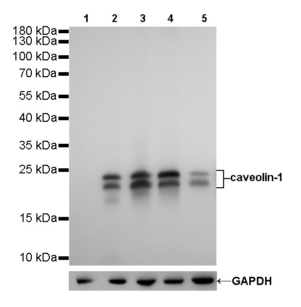
WB result of caveolin-1 Rabbit mAb
Primary antibody: caveolin-1 Rabbit mAb at 1/500 dilution
Lane 1: Daudi whole cell lysate 20 µg
Lane 2: PC-3 whole cell lysate 20 µg
Lane 3: A549 whole cell lysate 20 µg
Lane 4: A431 whole cell lysate 20 µg
Lane 5: HeLa whole cell lysate 20 µg
Negative control: Daudi whole cell lysate
Secondary antibody: Goat Anti-Rabbit IgG, (H+L), HRP conjugated at 1/10000 dilution
Predicted MW: 21, 24 kDa
Observed MW: 21,24 kDa
Exposure time: 6s
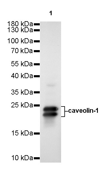
WB result of caveolin-1 Rabbit mAb
Primary antibody: caveolin-1 Rabbit mAb at 1/10000 dilution
Lane 1: U-87 MG whole cell lysate 20 µg
Secondary antibody: Goat Anti-Rabbit IgG, (H+L), HRP conjugated at 1/10000 dilution
Predicted MW: 21, 24 kDa
Observed MW: 21,24 kDa
Exposure time: 90s
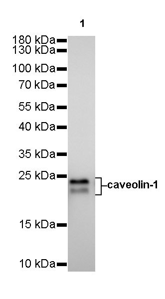
WB result of caveolin-1 Rabbit mAb
Primary antibody: caveolin-1 Rabbit mAb at 1/500 dilution
Lane 1: C2C12 whole cell lysate 20 µg
Secondary antibody: Goat Anti-Rabbit IgG, (H+L), HRP conjugated at 1/10000 dilution
Predicted MW: 21, 24 kDa
Observed MW: 21,24 kDa
Exposure time: 90s
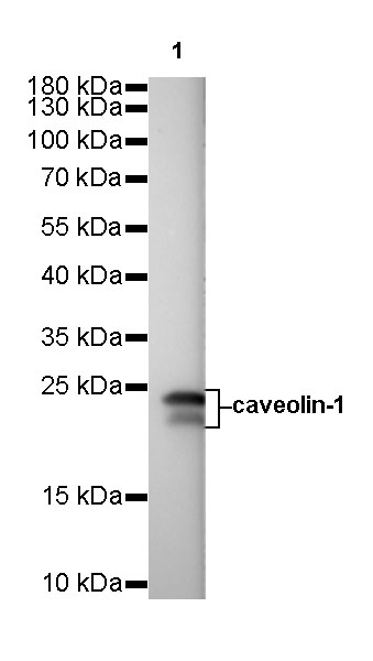
WB result of caveolin-1 Rabbit mAb
Primary antibody: caveolin-1 Rabbit mAb at 1/500 dilution
Lane 1: C6 whole cell lysate 20 µg
Secondary antibody: Goat Anti-Rabbit IgG, (H+L), HRP conjugated at 1/10000 dilution
Predicted MW: 21, 24 kDa
Observed MW: 21,24 kDa
Exposure time: 6s
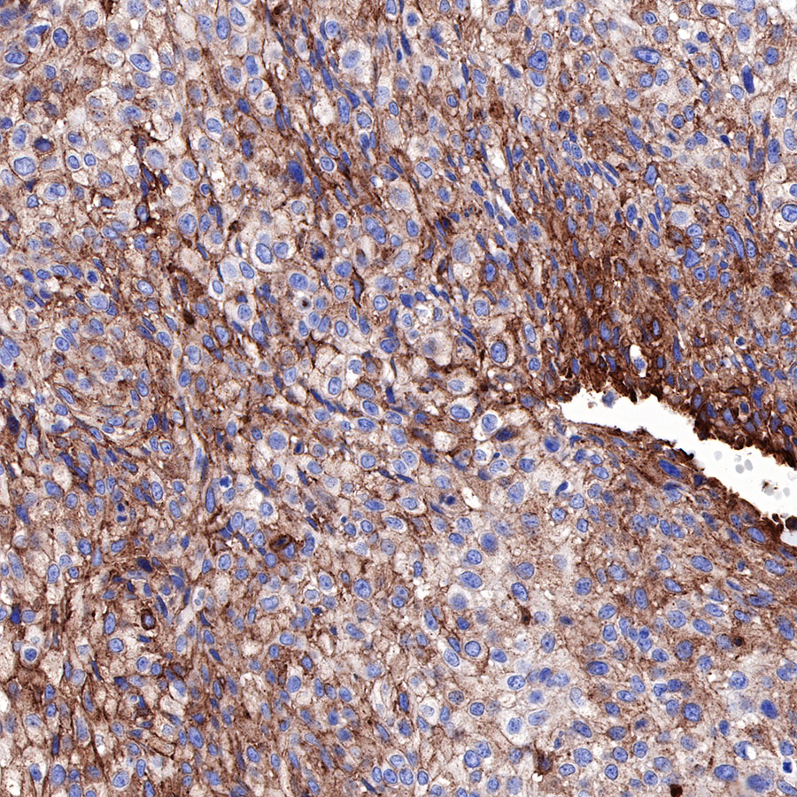
IHC shows positive staining in paraffin-embedded human cervial carcinoma. Anti-Caveolin-1 antibody was used at 1/1000 dilution, followed by a HRP Polymer for Mouse & Rabbit IgG (ready to use). Counterstained with hematoxylin. Heat mediated antigen retrieval with Tris/EDTA buffer pH9.0 was performed before commencing with IHC staining protocol.
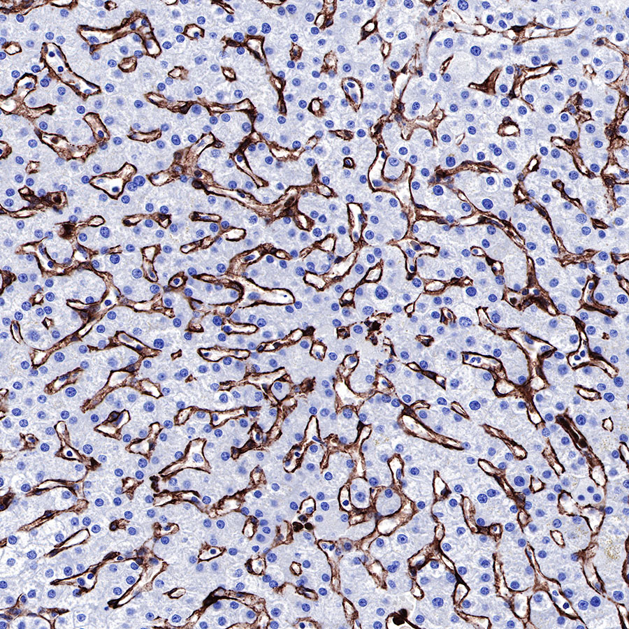
IHC shows positive staining in paraffin-embedded human liver. Anti-Caveolin-1 antibody was used at 1/1000 dilution, followed by a HRP Polymer for Mouse & Rabbit IgG (ready to use). Counterstained with hematoxylin. Heat mediated antigen retrieval with Tris/EDTA buffer pH9.0 was performed before commencing with IHC staining protocol.
IHC shows positive staining in paraffin-embedded human lung cancer. Anti-Caveolin-1 antibody was used at 1/1000 dilution, followed by a HRP Polymer for Mouse & Rabbit IgG (ready to use). Counterstained with hematoxylin. Heat mediated antigen retrieval with Tris/EDTA buffer pH9.0 was performed before commencing with IHC staining protocol.
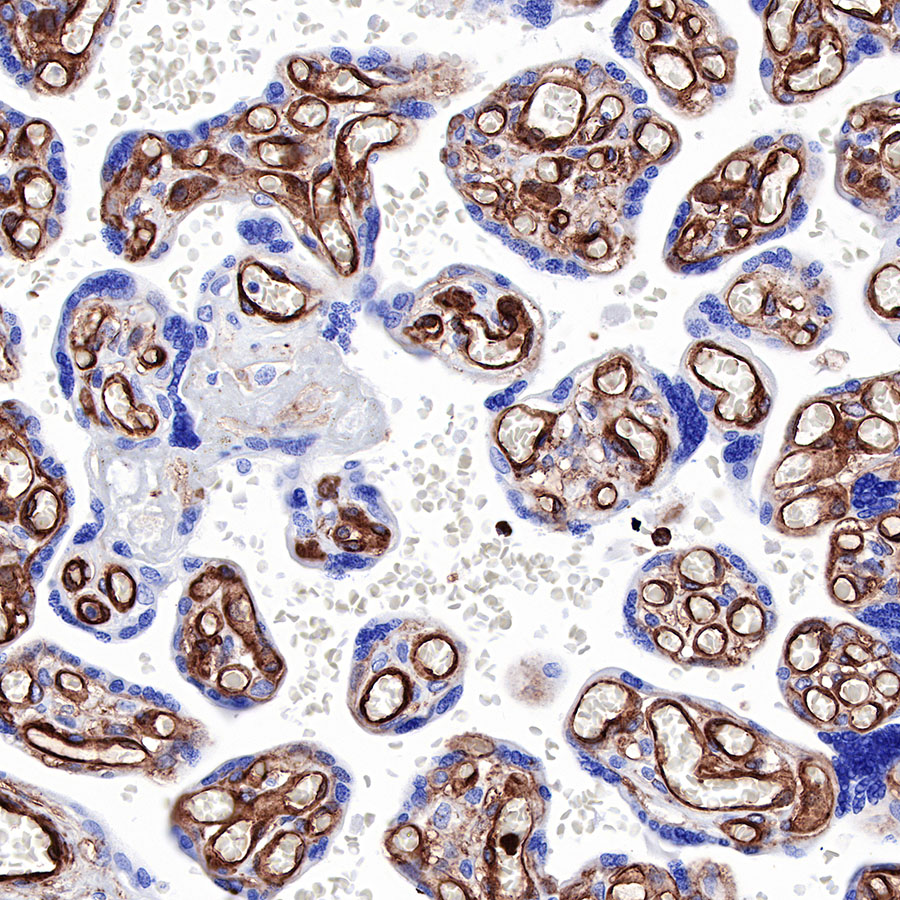
IHC shows positive staining in paraffin-embedded human placenta. Anti-Caveolin-1 antibody was used at 1/1000 dilution, followed by a HRP Polymer for Mouse & Rabbit IgG (ready to use). Counterstained with hematoxylin. Heat mediated antigen retrieval with Tris/EDTA buffer pH9.0 was performed before commencing with IHC staining protocol.
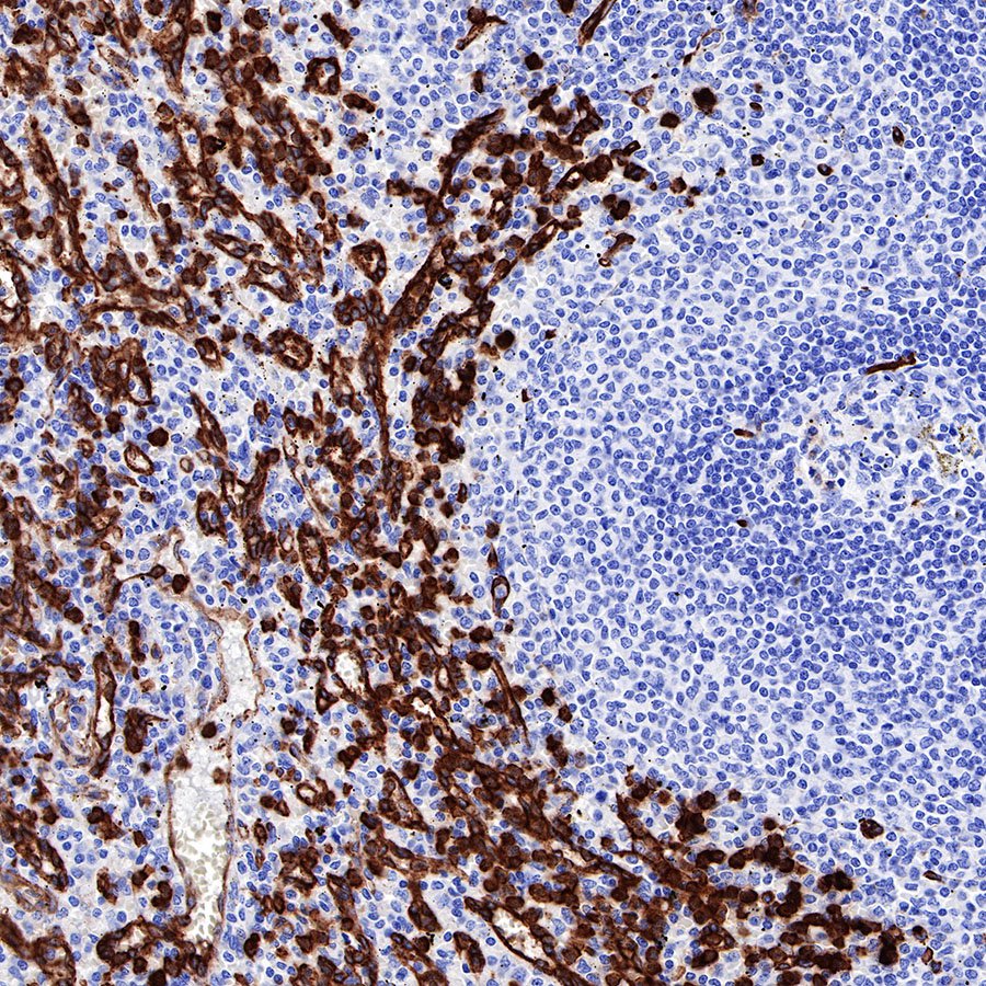
IHC shows positive staining in paraffin-embedded human spleen. Anti-Caveolin-1 antibody was used at 1/1000 dilution, followed by a HRP Polymer for Mouse & Rabbit IgG (ready to use). Counterstained with hematoxylin. Heat mediated antigen retrieval with Tris/EDTA buffer pH9.0 was performed before commencing with IHC staining protocol.
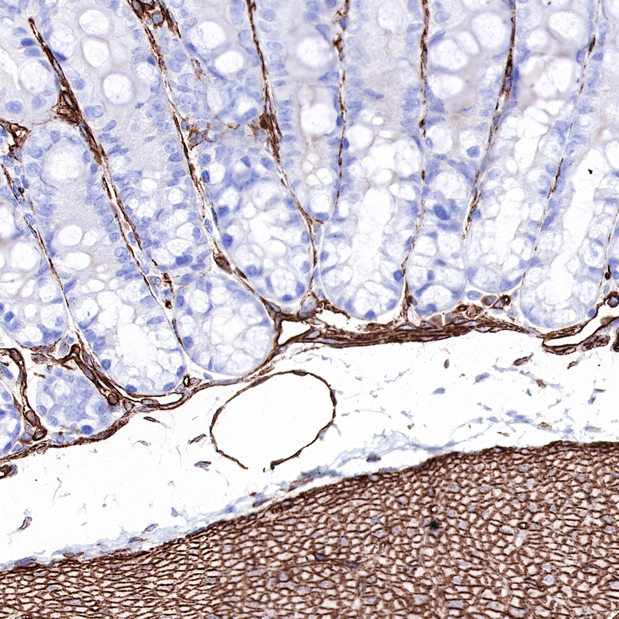
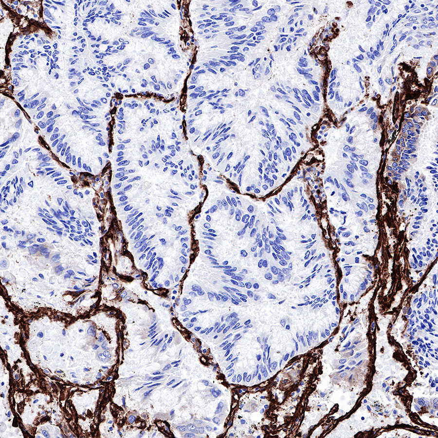
IHC shows positive staining in paraffin-embedded mouse colon. Anti-Caveolin-1 antibody was used at 1/1000 dilution, followed by a HRP Polymer for Mouse & Rabbit IgG (ready to use). Counterstained with hematoxylin. Heat mediated antigen retrieval with Tris/EDTA buffer pH9.0 was performed before commencing with IHC staining protocol.
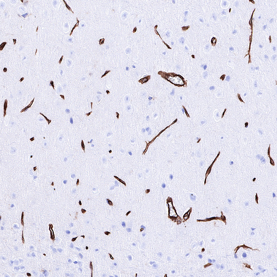
IHC shows positive staining in paraffin-embedded rat cerebral cortex. Anti-Caveolin-1 antibody was used at 1/1000 dilution, followed by a HRP Polymer for Mouse & Rabbit IgG (ready to use). Counterstained with hematoxylin. Heat mediated antigen retrieval with Tris/EDTA buffer pH9.0 was performed before commencing with IHC staining protocol.
 您现在的位置: