
PBS, 40% Glycerol, 0.05%BSA, 0.03% Proclin 300
12 months from date of receipt / reconstitution, -20 °C as supplied
| 应用 | 稀释度 |
|---|---|
| IHC-P | 1:500-1:1000 |
| ICFCM | 1:500 |
| WB | 1:1000 |
| ICC | 1:500 |
| IP | 1:25 |
The FOXP subfamily is defined by four members (FOXP1–FOXP4). FOXPs function as transcription factors and possess distinct transcriptional regulatory and DNA-binding domains. The N terminus of FOXPs contains a transcriptional repressor region with a zinc-finger/leucine zipper motif. The DNA-binding domain of FOXPs is uniquely positioned among forkhead proteins near the C terminus. FOXP1 is widely expressed in human tissues and regulates the development of many tissues, including heart, thymus, and lung. FOXP1-deficient embryos have severe defects in cardiac morphogenesis, including outflow tract septation and cushion defects, resulting in embryonic death.

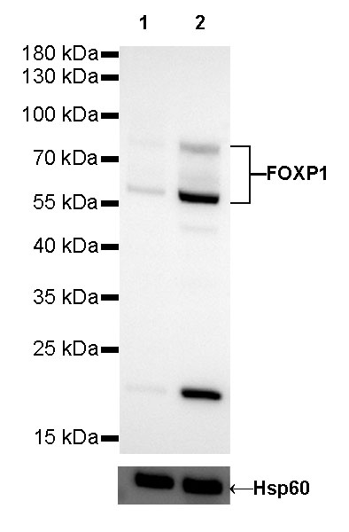
WB result of FOXP1 Rabbit mAb
Primary antibody: FOXP1 Rabbit mAb at 1/1000 dilution
Lane 1: A549 whole cell lysate 20 µg
Lane 2: Daudi whole cell lysate 20 µg
Low expression control: A549 whole cell lysate
Secondary antibody: Goat Anti-Rabbit IgG, (H+L), HRP conjugated at 1/10000 dilution
Predicted MW: 75 kDa
Observed MW: 60, 80 kDa
Exposure time: 180s
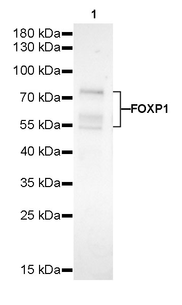
WB result of FOXP1 Rabbit mAb
Primary antibody: FOXP1 Rabbit mAb at 1/1000 dilution
Lane 1: mouse thymus lysate 20 µg
Secondary antibody: Goat Anti-Rabbit IgG, (H+L), HRP conjugated at 1/10000 dilution
Predicted MW: 75 kDa
Observed MW: 60, 80 kDa
Exposure time: 180s
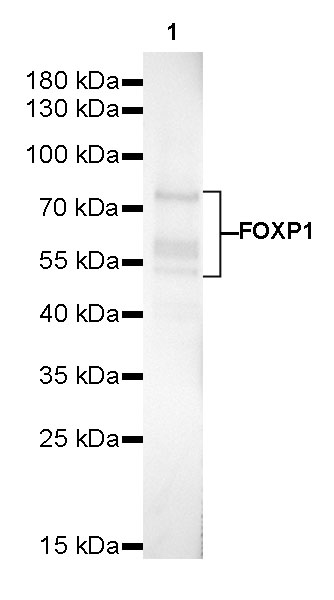
WB result of FOXP1 Rabbit mAb
Primary antibody: FOXP1 Rabbit mAb at 1/1000 dilution
Lane 1: rat thymus lysate 20 µg
Secondary antibody: Goat Anti-Rabbit IgG, (H+L), HRP conjugated at 1/10000 dilution
Predicted MW: 75 kDa
Observed MW: 60, 80 kDa
Exposure time: 180s
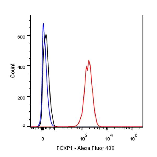
Flow cytometric analysis of Daudi cells labelling FOXP1 antibody at 1/500 (0.1 μg) dilution/ (red) compared with a Rabbit monoclonal IgG (Black) isotype control and an unlabelled control (cells without incubation with primary antibody and secondary antibody) (Blue). Goat Anti-Rabbit IgG Alexa Fluor® 488 was used as the secondary antibody.

FOXP1 Rabbit mAb at 1/25 dilution (2 µg) immunoprecipitating FOXP1 in 0.4 mg mouse thymus lysate.
Western blot was performed on the immunoprecipitate using FOXP1 Rabbit mAb at 1/1000 dilution.
Secondary antibody (HRP) for IP was used at 1/400 dilution.
Lane 1: mouse thymus lysate 20 µg (Input)
Lane 2: FOXP1 Rabbit mAb IP in mouse thymus lysate
Lane 3: Rabbit monoclonal IgG IP in mouse thymus lysate
Predicted MW: 75 kDa
Observed MW: 60, 80 kDa
Exposure time: 150 s
IHC shows positive staining in paraffin-embedded human breast. Anti-FOXP1 antibody was used at 1/500 dilution, followed by a HRP Polymer for Mouse & Rabbit IgG (ready to use). Counterstained with hematoxylin. Heat mediated antigen retrieval with Tris/EDTA buffer pH9.0 was performed before commencing with IHC staining protocol.
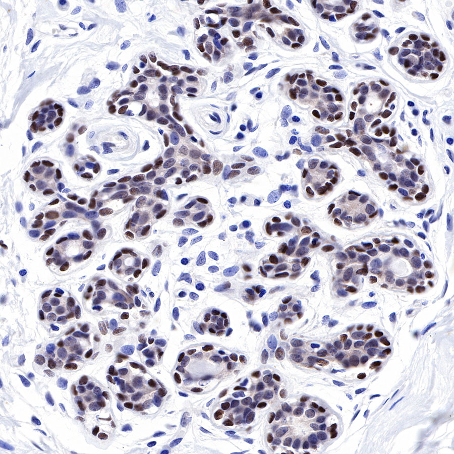
IHC shows positive staining in paraffin-embedded human breast cancer. Anti-FOXP1 antibody was used at 1/500 dilution, followed by a HRP Polymer for Mouse & Rabbit IgG (ready to use). Counterstained with hematoxylin. Heat mediated antigen retrieval with Tris/EDTA buffer pH9.0 was performed before commencing with IHC staining protocol.
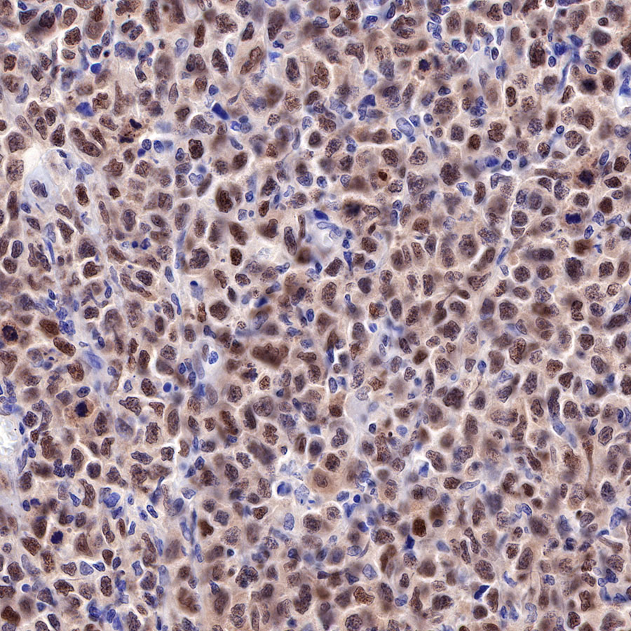
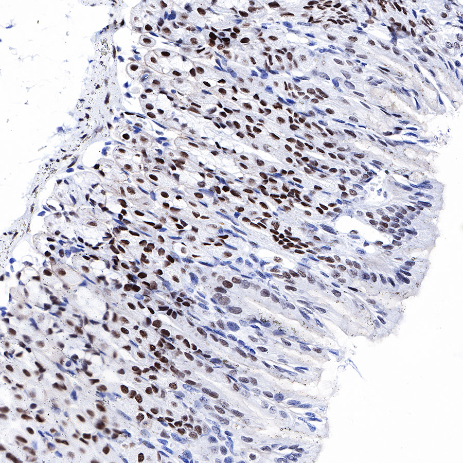
IHC shows positive staining in paraffin-embedded human diffuse large B-cell lymphoma. Anti-FOXP1 antibody was used at 1/500 dilution, followed by a HRP Polymer for Mouse & Rabbit IgG (ready to use). Counterstained with hematoxylin. Heat mediated antigen retrieval with Tris/EDTA buffer pH9.0 was performed before commencing with IHC staining protocol.
IHC shows positive staining in paraffin-embedded human colon. Anti-FOXP1 antibody was used at 1/500 dilution, followed by a HRP Polymer for Mouse & Rabbit IgG (ready to use). Counterstained with hematoxylin. Heat mediated antigen retrieval with Tris/EDTA buffer pH9.0 was performed before commencing with IHC staining protocol.
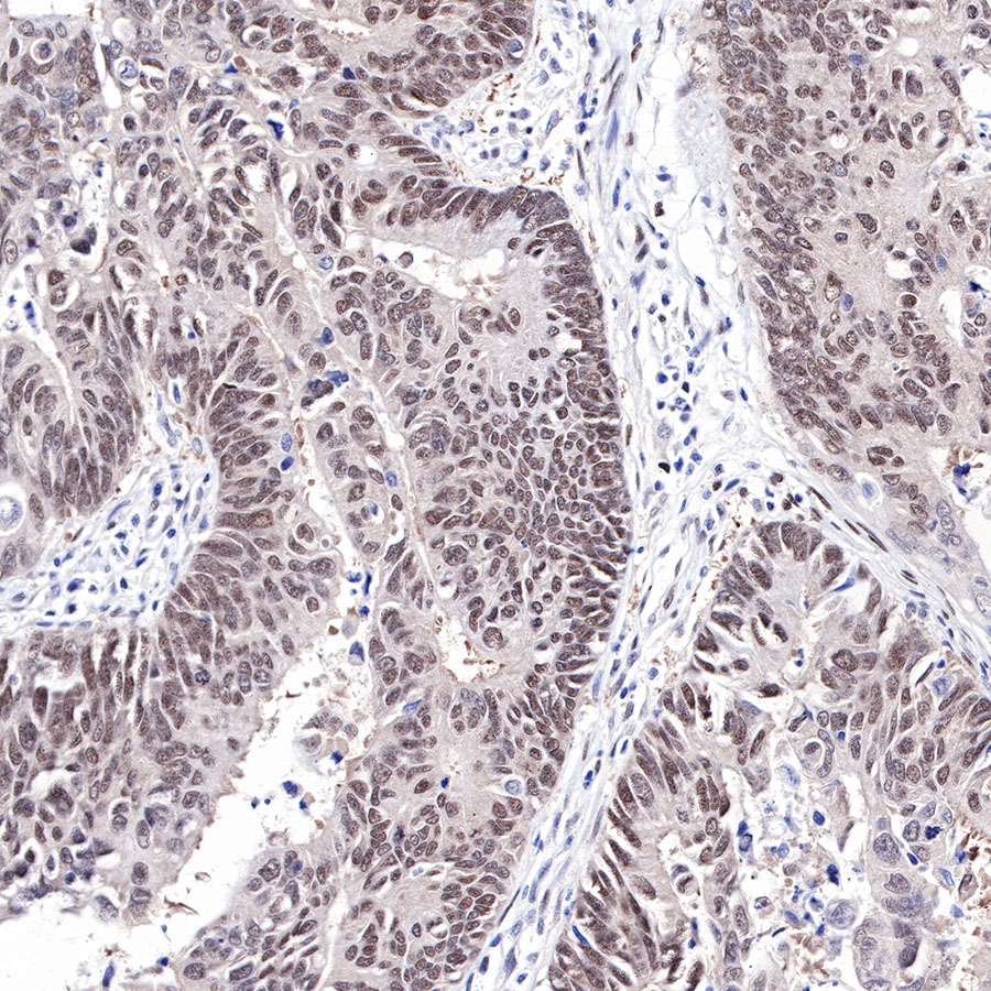
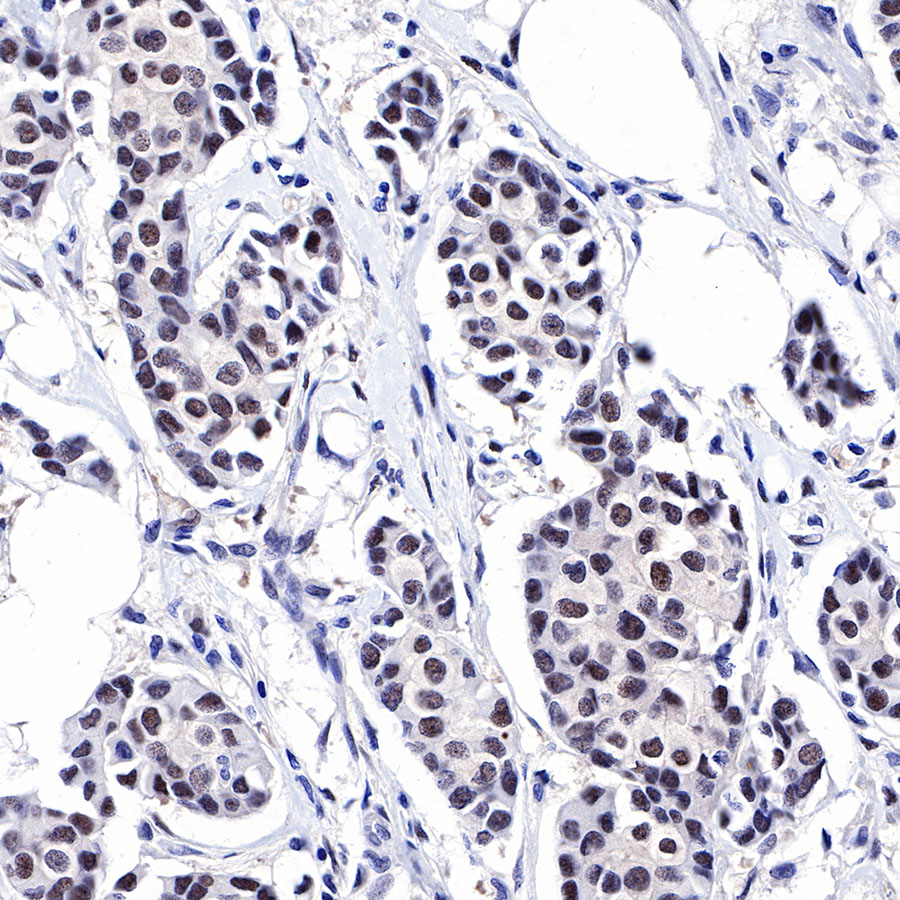
IHC shows positive staining in paraffin-embedded human colon cancer. Anti-FOXP1 antibody was used at 1/500 dilution, followed by a HRP Polymer for Mouse & Rabbit IgG (ready to use). Counterstained with hematoxylin. Heat mediated antigen retrieval with Tris/EDTA buffer pH9.0 was performed before commencing with IHC staining protocol.
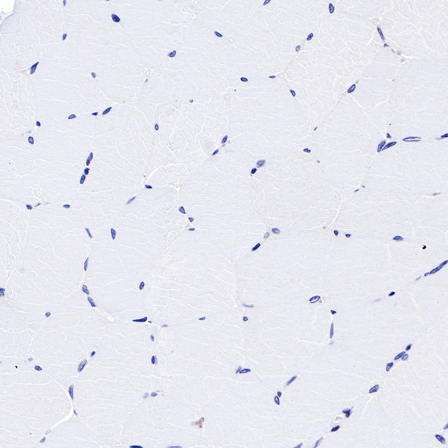
Negative control: IHC shows negative staining in paraffin-embedded human skeletal muscle. Anti-FOXP1 antibody was used at 1/500 dilution, followed by a HRP Polymer for Mouse & Rabbit IgG (ready to use). Counterstained with hematoxylin. Heat mediated antigen retrieval with Tris/EDTA buffer pH9.0 was performed before commencing with IHC staining protocol.
IHC shows positive staining in paraffin-embedded mouse stomach. Anti-FOXP1 antibody was used at 1/1000 dilution, followed by a HRP Polymer for Mouse & Rabbit IgG (ready to use). Counterstained with hematoxylin. Heat mediated antigen retrieval with Tris/EDTA buffer pH9.0 was performed before commencing with IHC staining protocol.

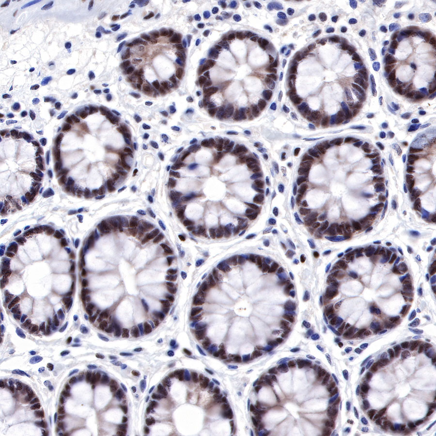
IHC shows positive staining in paraffin-embedded rat colon. Anti-FOXP1 antibody was used at 1/1000 dilution, followed by a HRP Polymer for Mouse & Rabbit IgG (ready to use). Counterstained with hematoxylin. Heat mediated antigen retrieval with Tris/EDTA buffer pH9.0 was performed before commencing with IHC staining protocol.

ICC shows positive staining in MCF7 cells. Anti- FOXP1 antibody was used at 1/500 dilution (Green) and incubated overnight at 4°C. Goat polyclonal Antibody to Rabbit IgG - H&L (Alexa Fluor® 488) was used as secondary antibody at 1/1000 dilution. The cells were fixed with 100% ice-cold methanol and permeabilized with 0.1% PBS-Triton X-100. Nuclei were counterstained with DAPI (Blue). Counterstain with tubulin (Red).
 您现在的位置: