

PBS, 40% Glycerol, 0.05%BSA, 0.03% Proclin 300
12 months from date of receipt / reconstitution, -20 °C as supplied
| 应用 | 稀释度 |
|---|---|
| FCM | 1:50 |
| WB | 1:1000 |
| IF | 1:1000 |
| IHC-P | 1:500 |
CD8A encodes the CD8 alpha chain of the αβT cells, proposed as a quantifiable indicator for CD8+ CTL recruitment or activity assessments and a robust biomarker for responses to anti-PD-1/PD-L1 therapy.In NK-cells, the presence of CD8A homodimers at the cell surface provides a survival mechanism allowing conjugation and lysis of multiple target cells. CD8A homodimer molecules also promote the survival and differentiation of activated lymphocytes into memory CD8 T-cells.
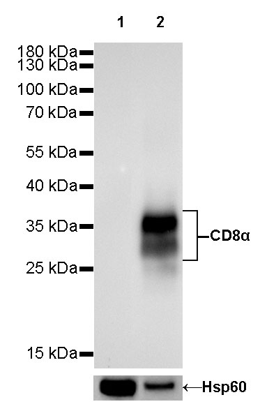
WB result of CD8α Rabbit mAb
Primary antibody: CD8α Rabbit mAb at 1/1000 dilution
Lane 1: RAW 264.7 whole cell lysate 20 µg
Lane 2: mouse thymus lysate 20 µg
Negative control: RAW 264.7 whole cell lysate
Secondary antibody: Goat Anti-Rabbit IgG, (H+L), HRP conjugated at 1/10000 dilution
Predicted MW: 29 kDa
Observed MW: 28~37 kDa
Exposure time: 60s
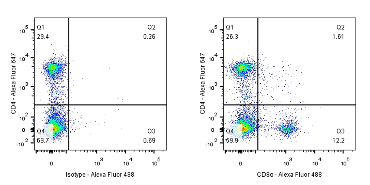
Flow cytometric analysis of mouse primary splenocytes labeling CD8α antibody at 1/50 (1 μg) dilution / (right panel) compared with a Rabbit IgG, Isotype Control / (left panel). Goat Anti-Rabbit IgG Alexa Fluor® 488 was used as the secondary antibody.
Cells were surface stained with CD4-Alexa Fluor® 647, then stained with rabbit IgG (Left) / CD8α (Right) separately. CD4 and CD8α are mutually exclusive expressed in mouse spleen. Gated on total viable cells.
IHC shows positive staining in paraffin-embedded mouse thymus. Anti-CD8α antibody was used at 1/500 dilution, followed by a HRP Polymer for Mouse & Rabbit IgG (ready to use). Counterstained with hematoxylin. Heat mediated antigen retrieval with Tris/EDTA buffer pH9.0 was performed before commencing with IHC staining protocol.
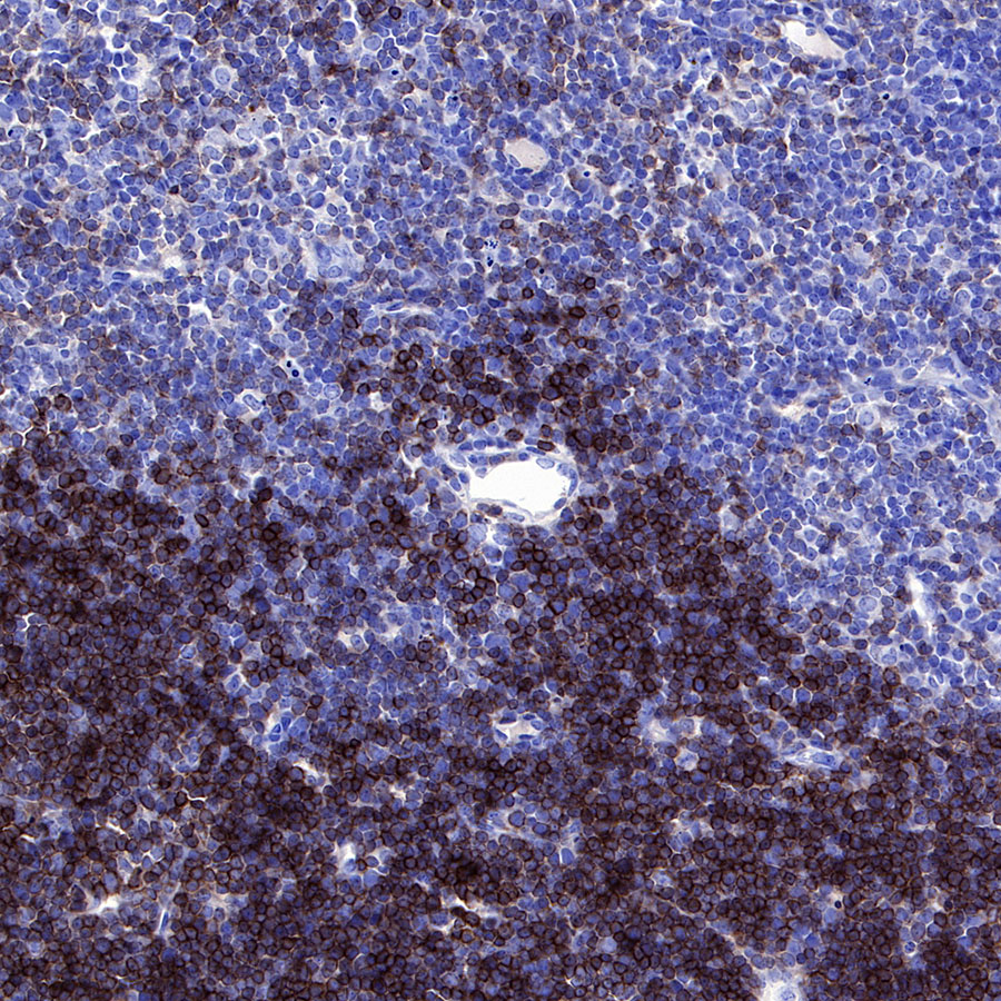
IHC shows positive staining in paraffin-embedded mouse spleen. Anti-CD8α antibody was used at 1/500 dilution, followed by a HRP Polymer for Mouse & Rabbit IgG (ready to use). Counterstained with hematoxylin. Heat mediated antigen retrieval with Tris/EDTA buffer pH9.0 was performed before commencing with IHC staining protocol.
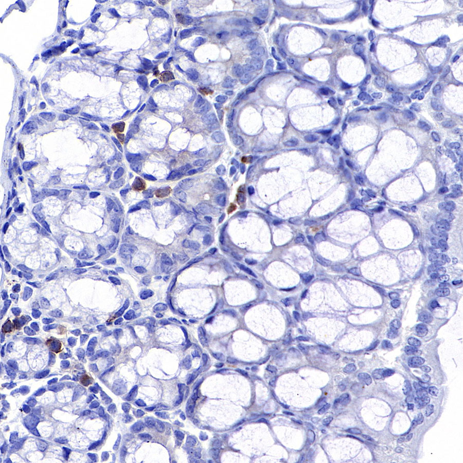
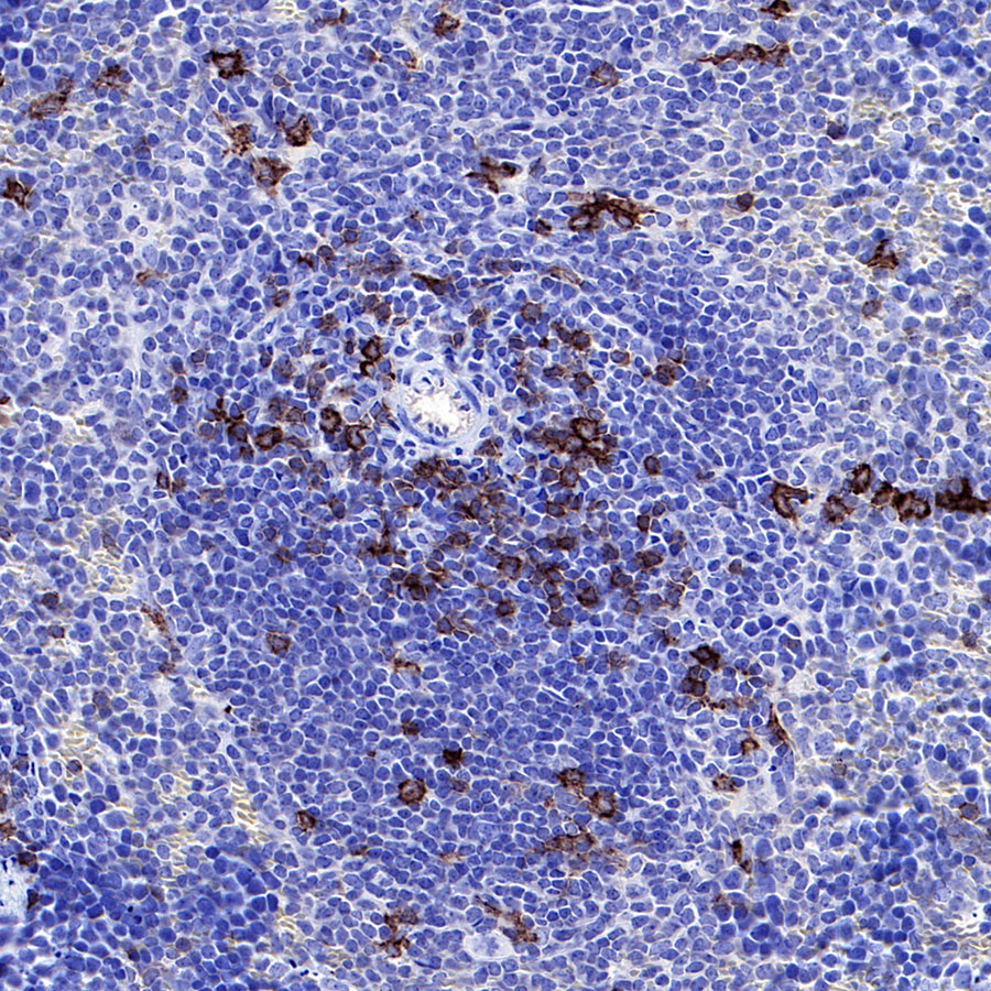
IHC shows positive staining in paraffin-embedded mouse colon. Anti-CD8α antibody was used at 1/500 dilution, followed by a HRP Polymer for Mouse & Rabbit IgG (ready to use). Counterstained with hematoxylin. Heat mediated antigen retrieval with Tris/EDTA buffer pH9.0 was performed before commencing with IHC staining protocol.
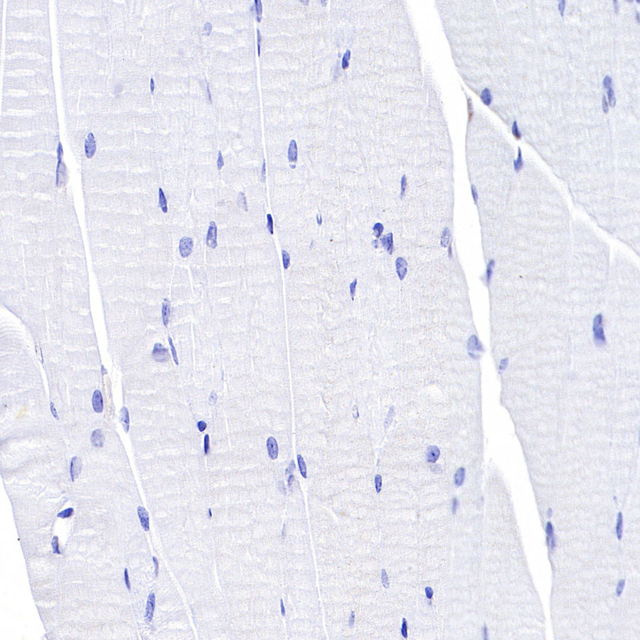
Negative control: IHC shows negative staining in paraffin-embedded mouse cardiac muscle. Anti-CD8α antibody was used at 1/500 dilution, followed by a HRP Polymer for Mouse & Rabbit IgG (ready to use). Counterstained with hematoxylin. Heat mediated antigen retrieval with Tris/EDTA buffer pH9.0 was performed before commencing with IHC staining protocol.
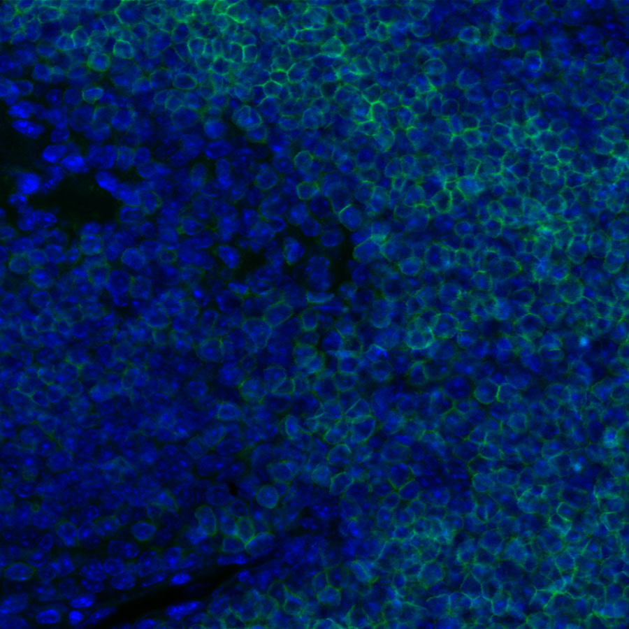
IF shows positive staining in paraffin-embedded mouse thymus. Anti-CD8α antibody was used at 1/1000 dilution and incubated overnight at 4°C. Goat polyclonal Antibody to Rabbit IgG - H&L (Alexa Fluor® 488) was used as secondary antibody at 1/1000 dilution.Heat mediated antigen retrieval with Tris/EDTA buffer pH9.0 was performed before commencing with ICC staining protocol. Nuclei were counterstained with DAPI.
 您现在的位置: