
PBS, 40% Glycerol, 0.05%BSA, 0.03% Proclin 300
12 months from date of receipt / reconstitution, -20 °C as supplied
| 应用 | 稀释度 |
|---|---|
| WB | 1:500-1:1000 |
| IHC-P | 1:1000 |
| IP | 1:25 |
| ICC | 1:250 |
Hepatocarcinoma is one of the most prevalent gastroenterological cancers in the world with less effective therapy. As an oncofetal antigen and diagnostic marker for liver cancer, alpha-fetoprotein (AFP) possesses a variety of biological functions. Except for its diagnosis in liver cancer, AFP has become a target for liver cancer immunotherapy. Although the immunogenicity of AFP is weak and it could induce the immune escapes through inhibiting the function of dendritic cells, natural killer cells, and T lymphocytes, AFP has attracted more attention in liver cancer immunotherapy.

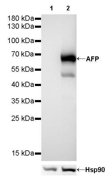
WB result of AFP Rabbit mAb
Primary antibody: AFP Rabbit mAb at 1/500 dilution
Lane 1: HeLa whole cell lysate 20 µg
Lane 2: HepG2 whole cell lysate 20 µg
Negative control: HeLa whole cell lysate
Secondary antibody: Goat Anti-Rabbit IgG, (H+L), HRP conjugated at 1/10000 dilution
Predicted MW: 69 kDa
Observed MW: 69 kDa
Exposure time: 30s
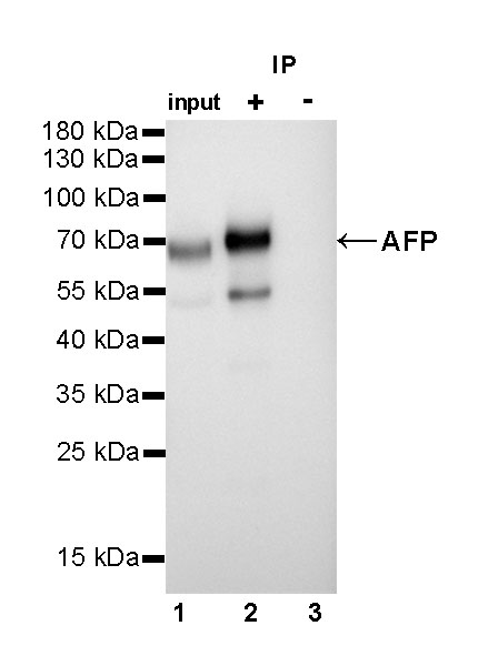
AFP Rabbit mAb at 1/25 dilution (1µg) immunoprecipitating AFP in 0.4mg HepG2 whole cell lysate.
Western blot was performed on the immunoprecipitate using AFP Rabbit mAb at 1/2000 dilution.
Secondary antibody (HRP) for IP was used at 1/400 dilution.
Lane 1 : HepG2 whole cell lysate 10µg (input)
Lane 2 : AFP Rabbit mAb IP in HepG2 whole cell lysate
Lane 3 : Rabbit monoclonal IgG IP in HepG2 whole cell lysate
Predicted MW: 69 kDa
Observed MW: 69 kDa
Exposure time: 5s
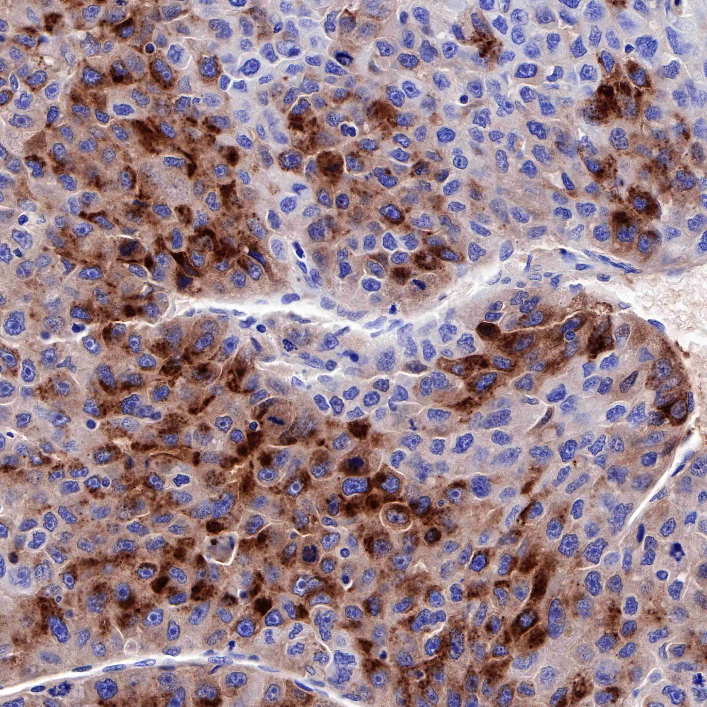
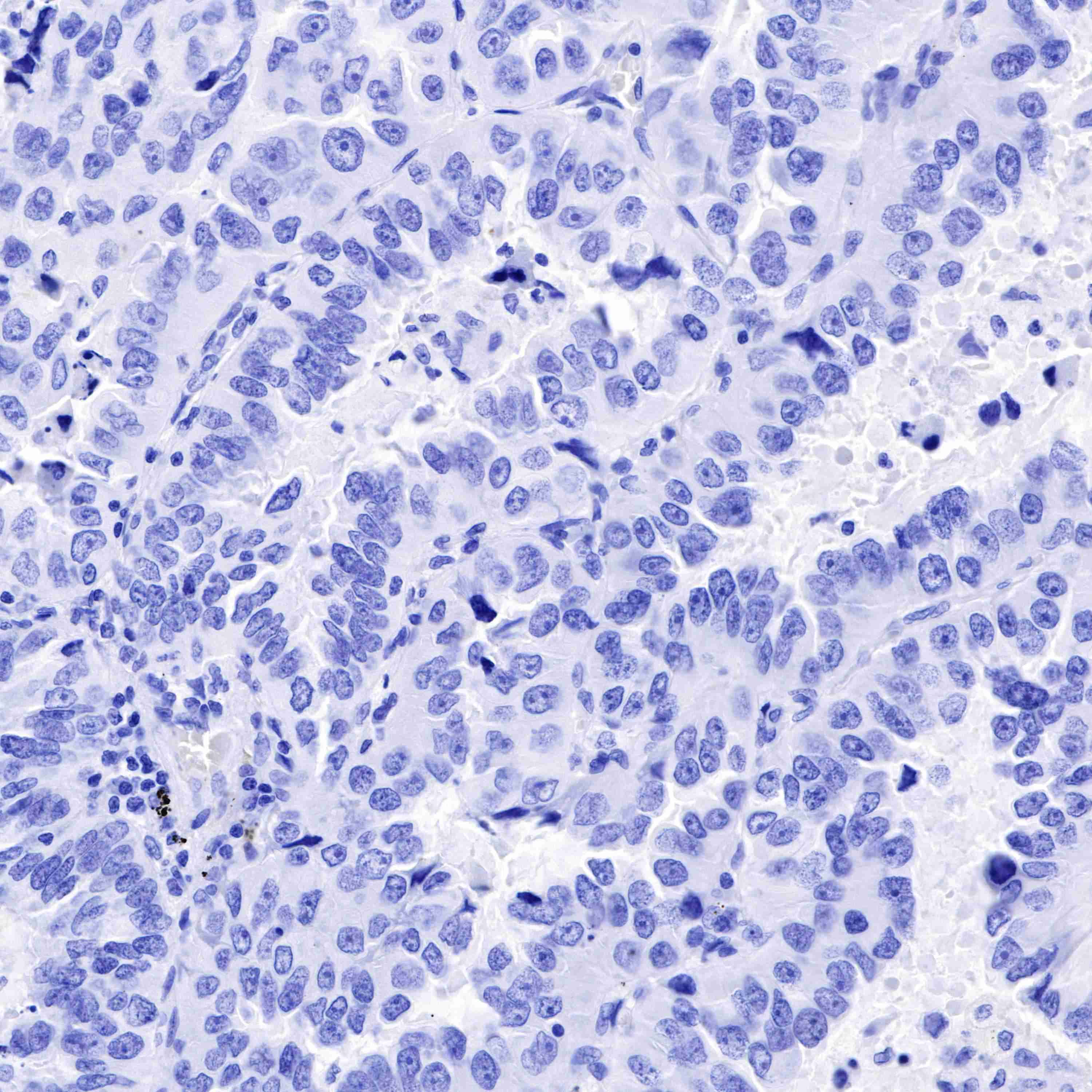
IHC shows positive staining in paraffin-embedded human hepatocellular carcinoma. Anti-AFP antibody was used at 1/1000 dilution, followed by a HRP Polymer for Mouse & Rabbit IgG (ready to use). Counterstained with hematoxylin. Heat mediated antigen retrieval with Tris/EDTA buffer pH9.0 was performed before commencing with IHC staining protocol.
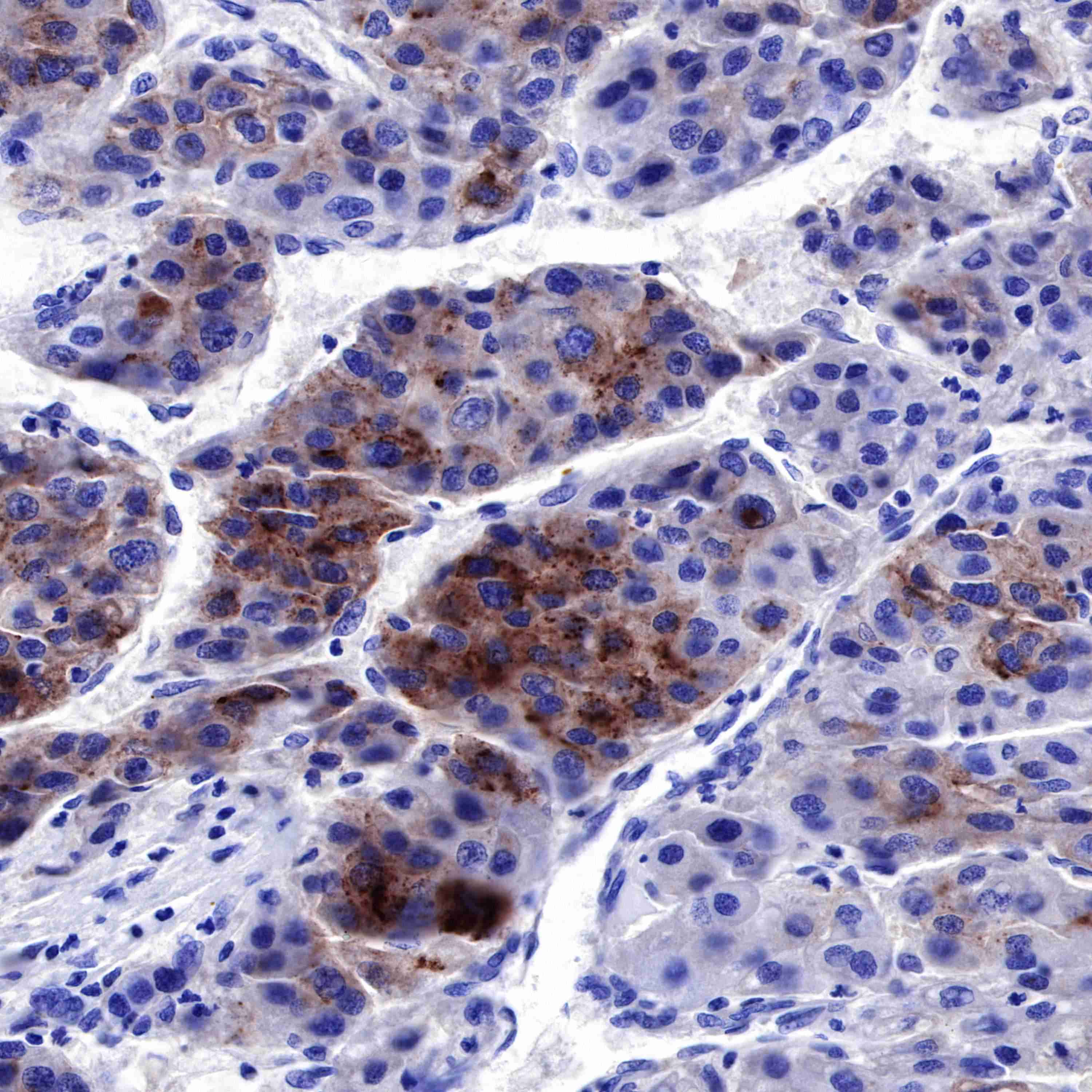
IHC shows positive staining in paraffin-embedded human hepatocellular carcinoma. Anti-AFP antibody was used at 1/1000 dilution, followed by a HRP Polymer for Mouse & Rabbit IgG (ready to use). Counterstained with hematoxylin. Heat mediated antigen retrieval with Tris/EDTA buffer pH9.0 was performed before commencing with IHC staining protocol.
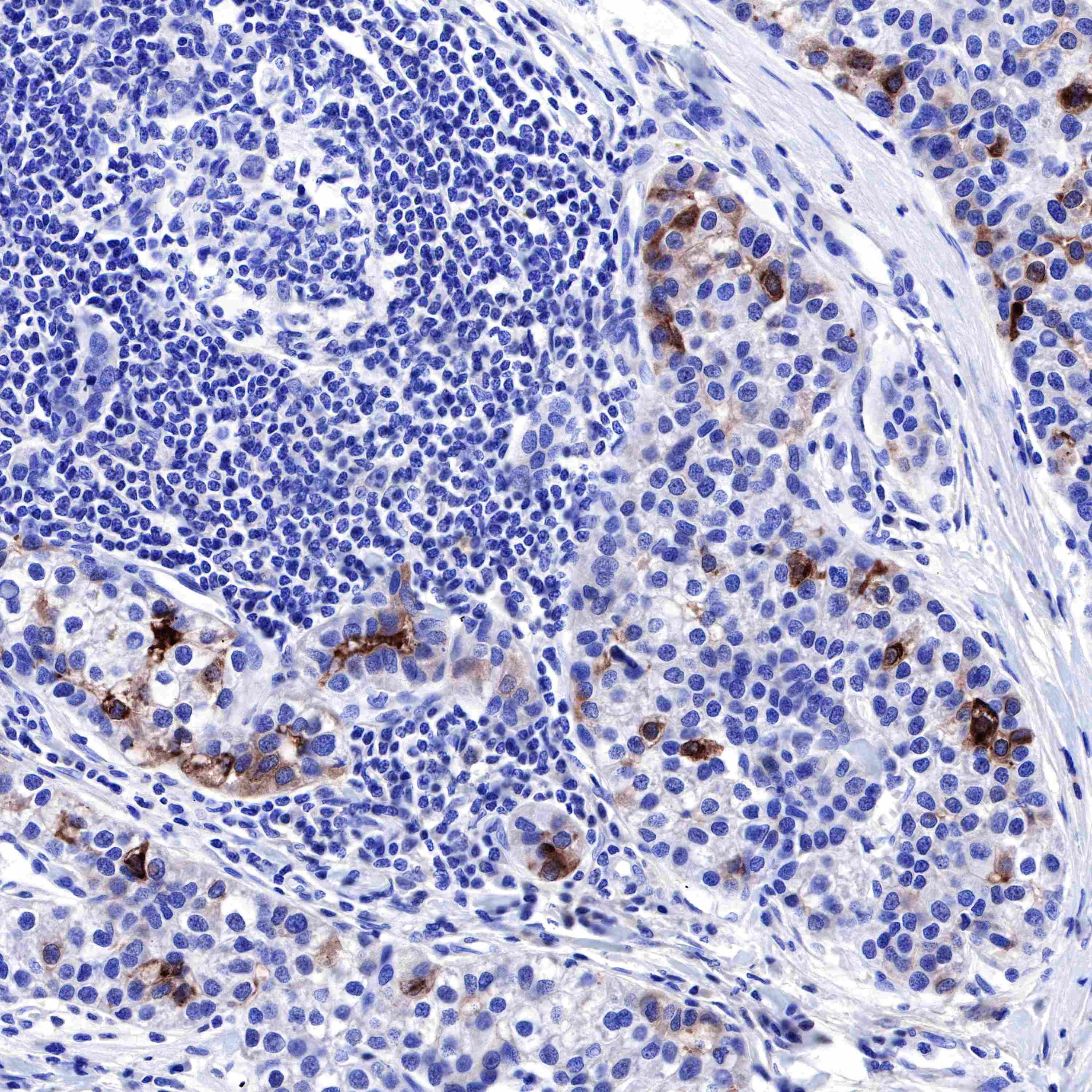
IHC shows positive staining in paraffin-embedded human hepatocellular carcinoma. Anti-AFP antibody was used at 1/1000 dilution, followed by a HRP Polymer for Mouse & Rabbit IgG (ready to use). Counterstained with hematoxylin. Heat mediated antigen retrieval with Tris/EDTA buffer pH9.0 was performed before commencing with IHC staining protocol.
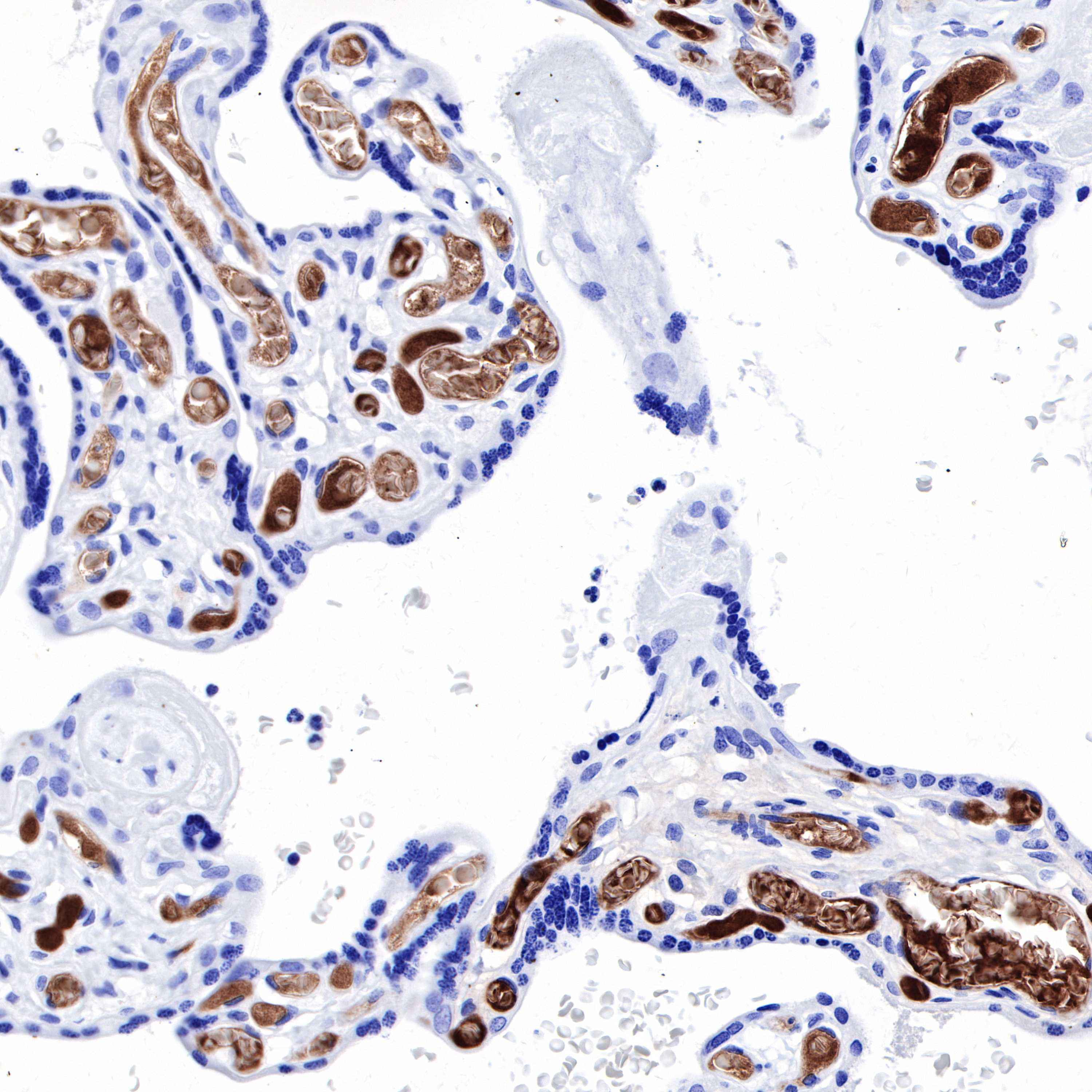
IHC shows positive staining in paraffin-embedded human placenta. Anti-AFP antibody was used at 1/1000 dilution, followed by a HRP Polymer for Mouse & Rabbit IgG (ready to use). Counterstained with hematoxylin. Heat mediated antigen retrieval with Tris/EDTA buffer pH9.0 was performed before commencing with IHC staining protocol.
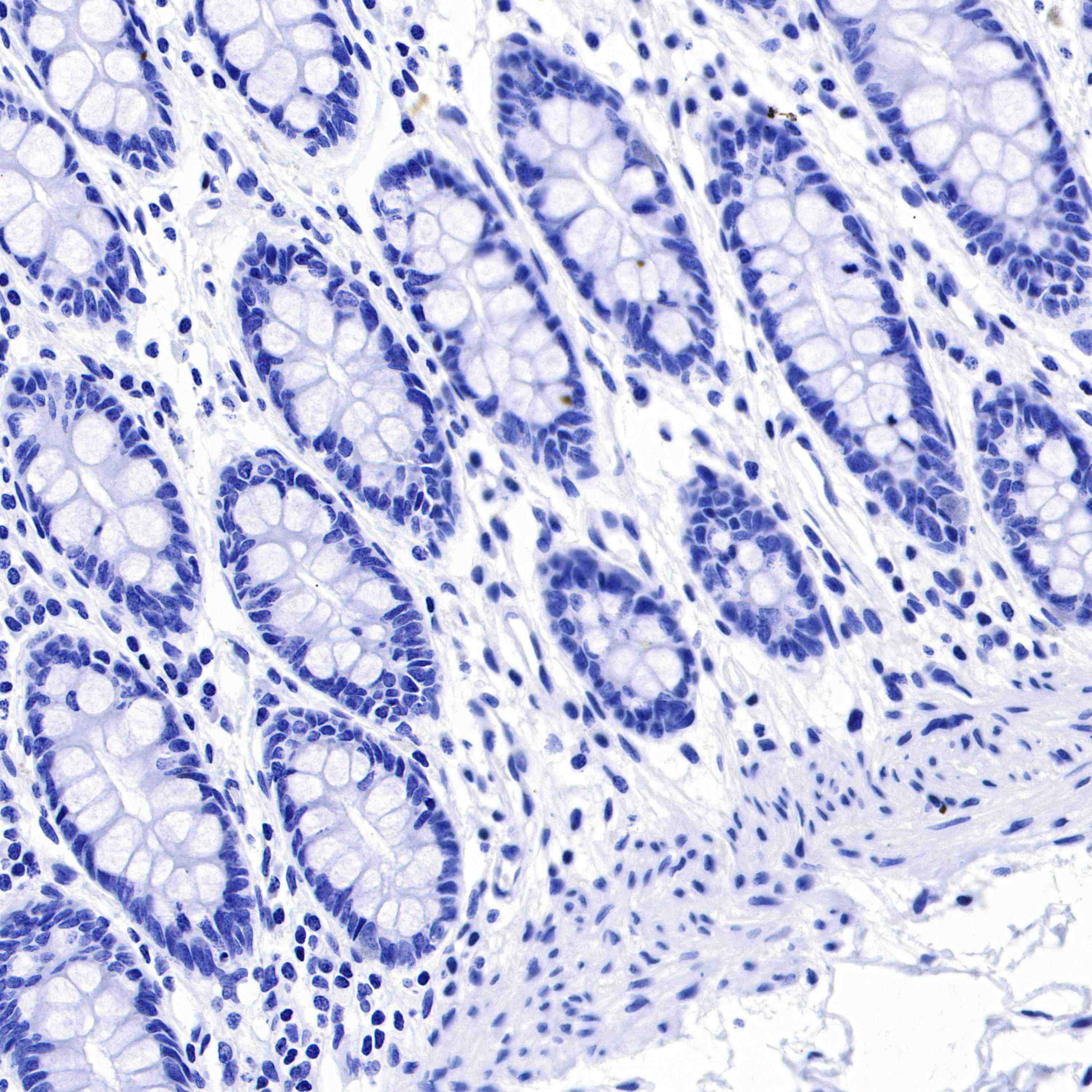
Negative control: IHC shows negative staining in paraffin-embedded human colon. Anti-AFP antibody was used at 1/1000 dilution, followed by a HRP Polymer for Mouse & Rabbit IgG (ready to use). Counterstained with hematoxylin. Heat mediated antigen retrieval with Tris/EDTA buffer pH9.0 was performed before commencing with IHC staining protocol.
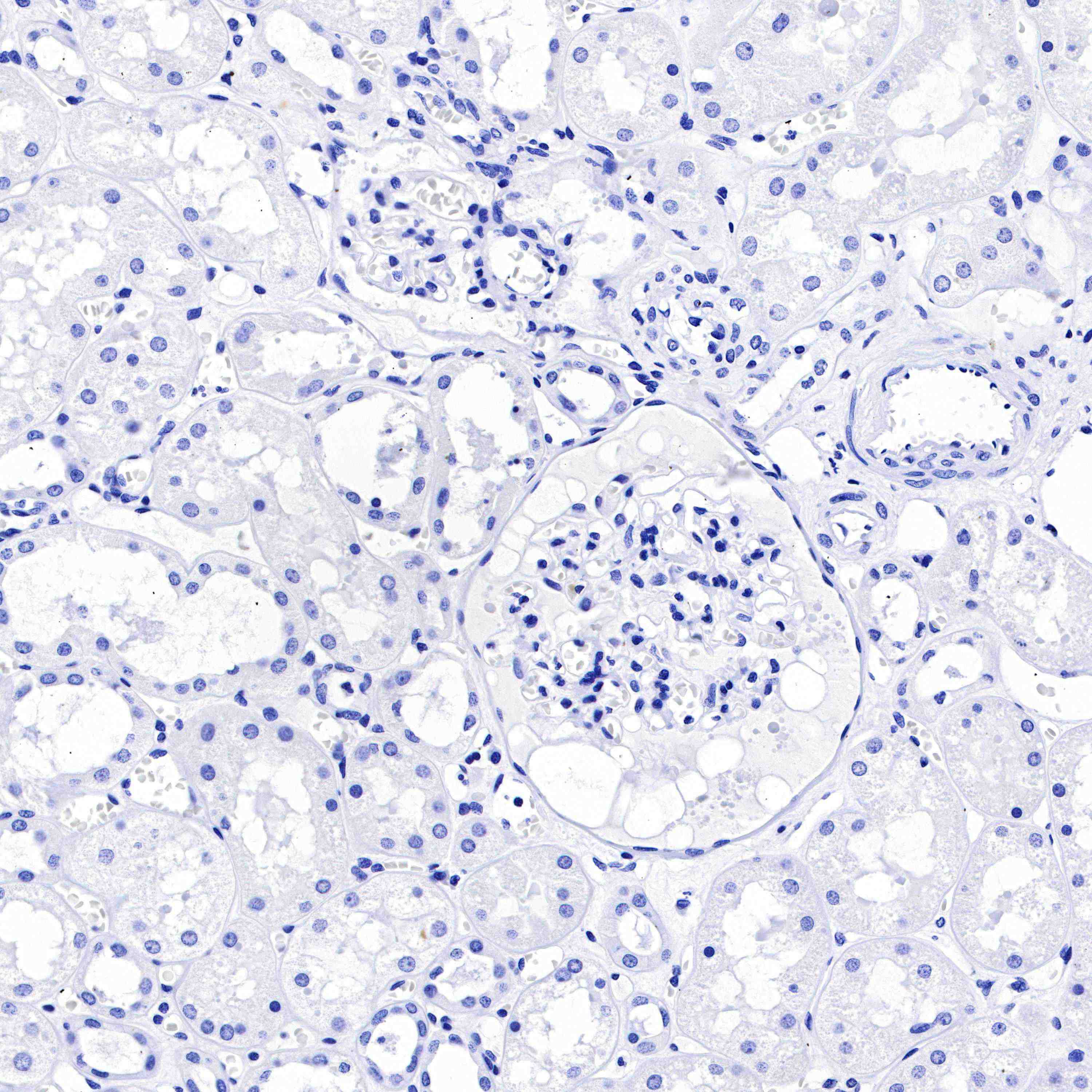
Negative control: IHC shows negative staining in paraffin-embedded human kidney. Anti-AFP antibody was used at 1/1000 dilution, followed by a HRP Polymer for Mouse & Rabbit IgG (ready to use). Counterstained with hematoxylin. Heat mediated antigen retrieval with Tris/EDTA buffer pH9.0 was performed before commencing with IHC staining protocol.
Negative control: IHC shows negative staining in paraffin-embedded human lung adenocarcinoma. Anti-AFP antibody was used at 1/1000 dilution, followed by a HRP Polymer for Mouse & Rabbit IgG (ready to use). Counterstained with hematoxylin. Heat mediated antigen retrieval with Tris/EDTA buffer pH9.0 was performed before commencing with IHC staining protocol.
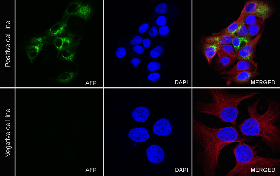
ICC shows positive staining in HepG2 cells (top panel) and negative staining in HeLa cells (below panel). Anti-AFP antibody was used at 1/250 dilution (Green) and incubated overnight at 4°C. Goat polyclonal Antibody to Rabbit IgG - H&L (Alexa Fluor® 488) was used as secondary antibody at 1/1000 dilution. The cells were fixed with 4% PFA and permeabilized with 0.1% PBS-Triton X-100. Nuclei were counterstained with DAPI (Blue). Counterstain with tubulin (Red).
 您现在的位置: