

| 应用 | 稀释度 |
|---|---|
| WB | 1:1000-1:2000 |
| IHC-P | 1:1000 |
| ICC | 1:125 |
SATB2, a nuclear protein known to bind matrix attachment regions, is a key event in initiating myogenic differentiation. The deletion of myoblast SATB2 in vitro initiates chromatin remodeling and accelerates differentiation, which is dependent on the caspase 7-mediated cleavage of SATB2. A genome-wide analysis indicates that SATB2 binding within chromatin loops and near anchor points influences both loop and sub-TAD domain formation. Consequently, the chromatin changes that occur with the removal of SATB2 lead to the derepression of differentiation-inducing factors while also limiting the expression of genes that inhibit this cell fate change.
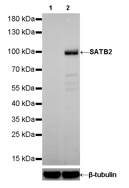
WB result of SATB2 Rabbit mAb
Primary antibody:SATB2 Rabbit mAb at 1/1250 dilution
Lane 1: SH-SY5Y whole cell lysate 20 µg
Lane 2: K-562 whole cell lysate 20 µg
Negative control: SH-SY5Y whole cell lysate
Secondary antibody: Goat Anti-Rabbit IgG, (H+L), HRP conjugated at 1/10000 dilution
Predicted MW: 100 kDa
Observed MW: 100 kDa

IHC shows positive staining in paraffin-embedded human cerebral cortex. Anti-SATB2 antibody was used at 1/1000 dilution, followed by a HRP Polymer for Mouse & Rabbit IgG (ready to use). Counterstained with hematoxylin. Heat mediated antigen retrieval with Tris/EDTA buffer pH9.0 was performed before commencing with IHC staining protocol.
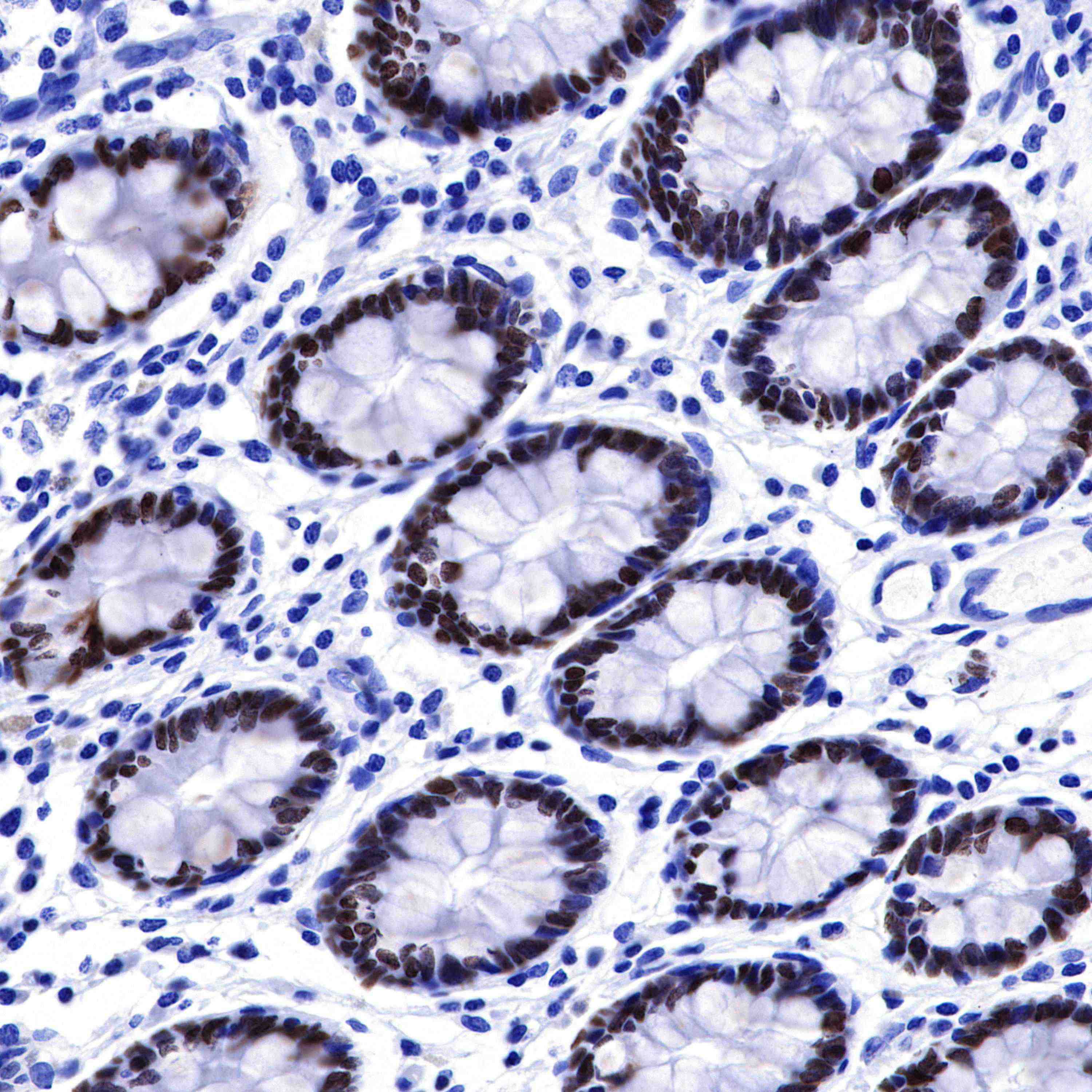
IHC shows positive staining in paraffin-embedded human colon. Anti-SATB2 antibody was used at 1/1000 dilution, followed by a HRP Polymer for Mouse & Rabbit IgG (ready to use). Counterstained with hematoxylin. Heat mediated antigen retrieval with Tris/EDTA buffer pH9.0 was performed before commencing with IHC staining protocol.
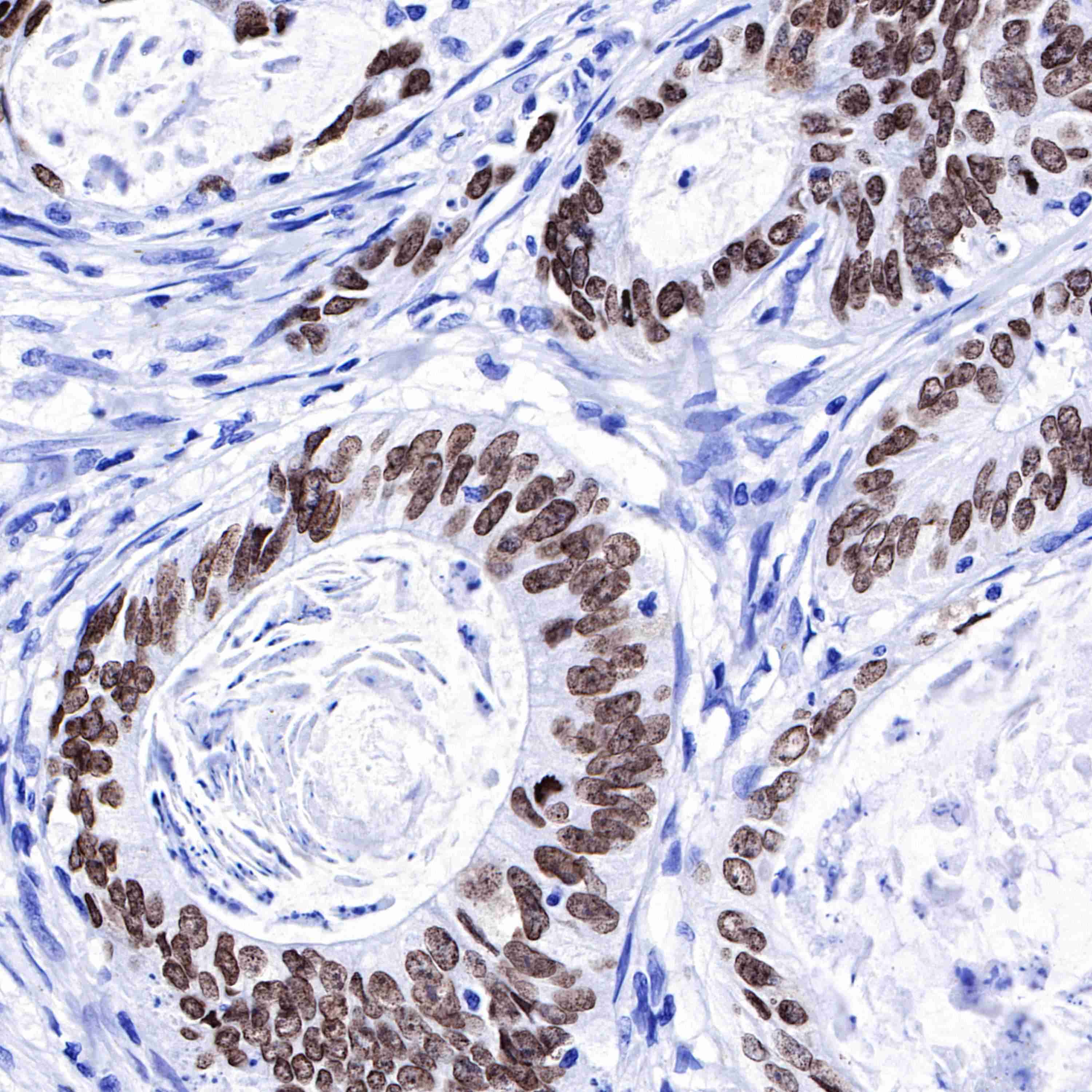
IHC shows positive staining in paraffin-embedded human colon cancer. Anti-SATB2 antibody was used at 1/1000 dilution, followed by a HRP Polymer for Mouse & Rabbit IgG (ready to use). Counterstained with hematoxylin. Heat mediated antigen retrieval with Tris/EDTA buffer pH9.0 was performed before commencing with IHC staining protocol.
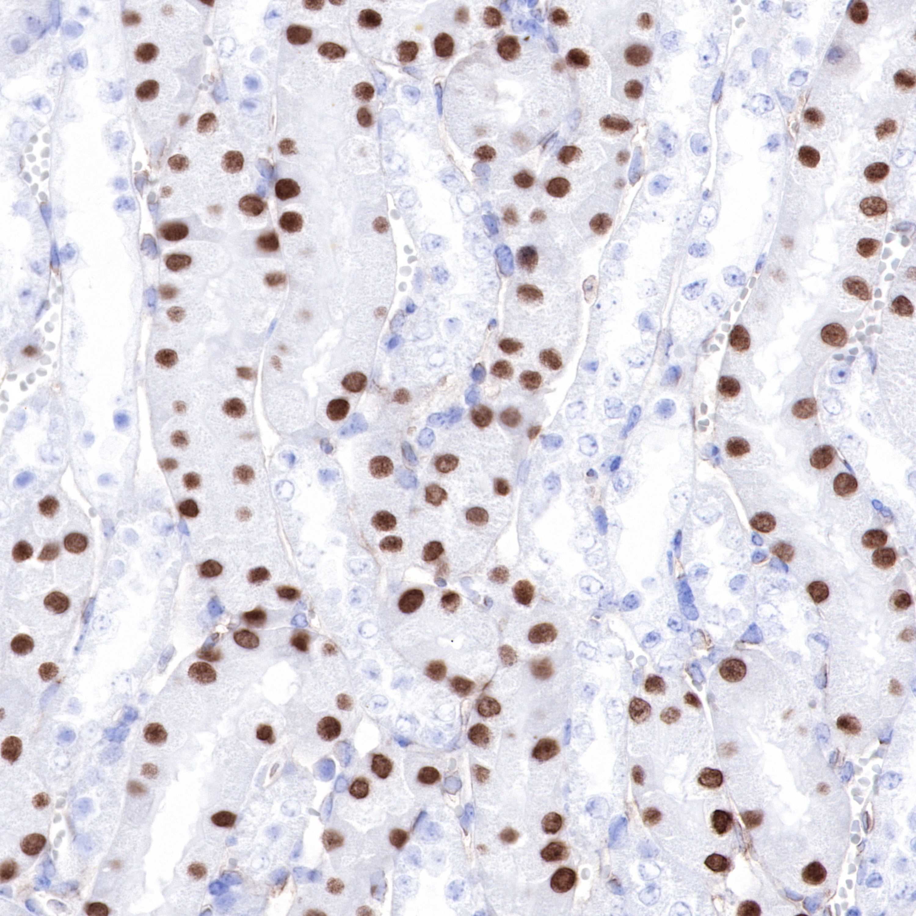
IHC shows positive staining in paraffin-embedded mouse kidney. Anti-SATB2 antibody was used at 1/1000 dilution, followed by a HRP Polymer for Mouse & Rabbit IgG (ready to use). Counterstained with hematoxylin. Heat mediated antigen retrieval with Tris/EDTA buffer pH9.0 was performed before commencing with IHC staining protocol.
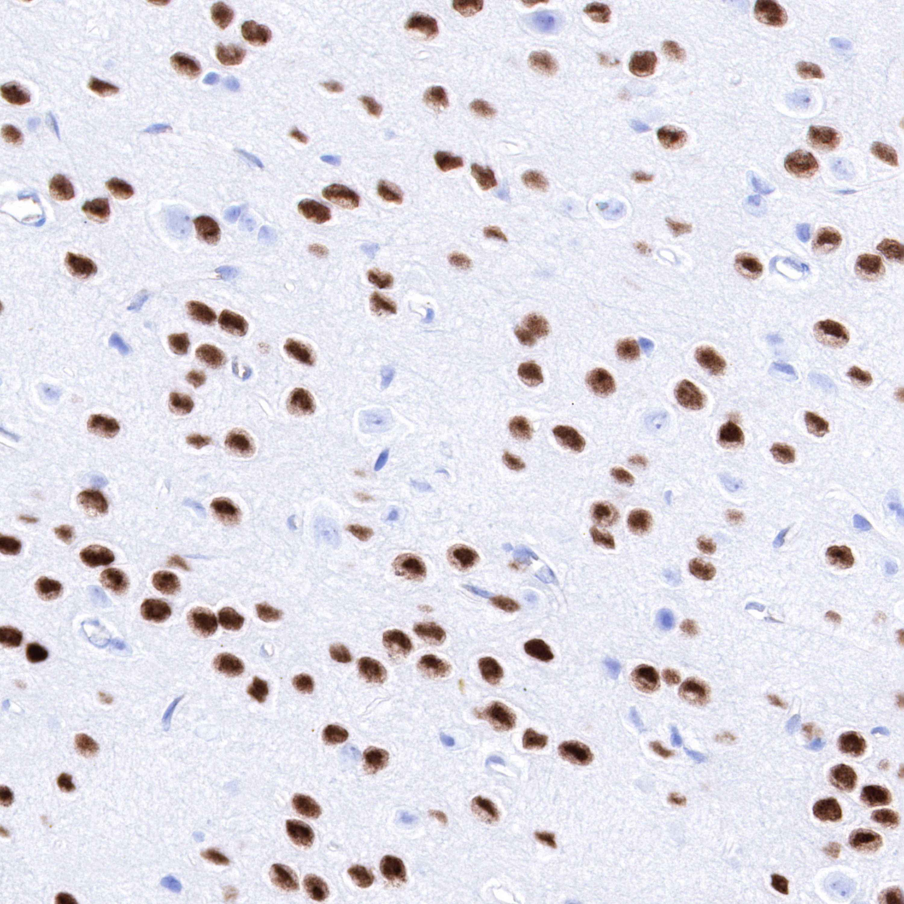
IHC shows positive staining in paraffin-embedded rat cerebral cortex. Anti-SATB2 antibody was used at 1/1000 dilution, followed by a HRP Polymer for Mouse & Rabbit IgG (ready to use). Counterstained with hematoxylin. Heat mediated antigen retrieval with Tris/EDTA buffer pH9.0 was performed before commencing with IHC staining protocol.
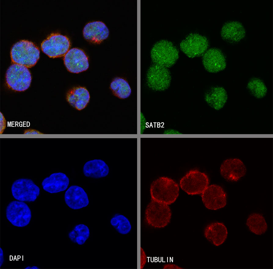
ICC shows positive staining in K562 cells. Anti-SATB2 antibody was used at 1/125 dilution (Green) and incubated overnight at 4°C. Goat polyclonal Antibody to Rabbit IgG - H&L (Alexa Fluor® 488) was used as secondary antibody at 1/1000 dilution. The cells were fixed with 4% PFA and permeabilized with 0.1% PBS-Triton X-100. Nuclei were counterstained with DAPI (Blue). Counterstain with tubulin (red).
 您现在的位置: