
PBS, 40% Glycerol, 0.05%BSA, 0.03% Proclin 300
12 months from date of receipt / reconstitution, -20 °C as supplied
| 应用 | 稀释度 |
|---|---|
| IHC-P | 1:1000 |
| WB | 1:1000-1:10000 |
| ICFCM | 1:500 |
| IP | 1:25 |
| ICC | 1:500 |
Human distal upstream element (Fuse) binding protein 1 (FUBP1) is a transcriptional regulator of c-Myc and represents an important prognostic marker in many cancers.Regulates MYC expression by binding to a single-stranded far-upstream element (FUSE) upstream of the MYC promoter. May act both as activator and repressor of transcription.

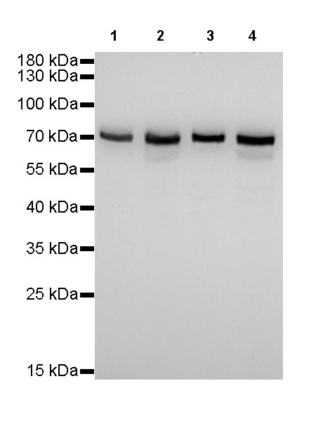
WB result of FUBP1 Rabbit mAb
Primary antibody: FUBP1 Rabbit mAb at 1/1000 dilution
Lane 1: HEK293 whole cell lysate 20 µg
Lane 2: Jurkat whole cell lysate 20 µg
Lane 3: HeLa whole cell lysate 20 µg
Lane 4: Raji whole cell lysate 20 µg
Secondary antibody: Goat Anti-Rabbit IgG, (H+L), HRP conjugated at 1/10000 dilution
Predicted MW: 67 kDa
Observed MW: 67 kDa
Exposure time: 9s
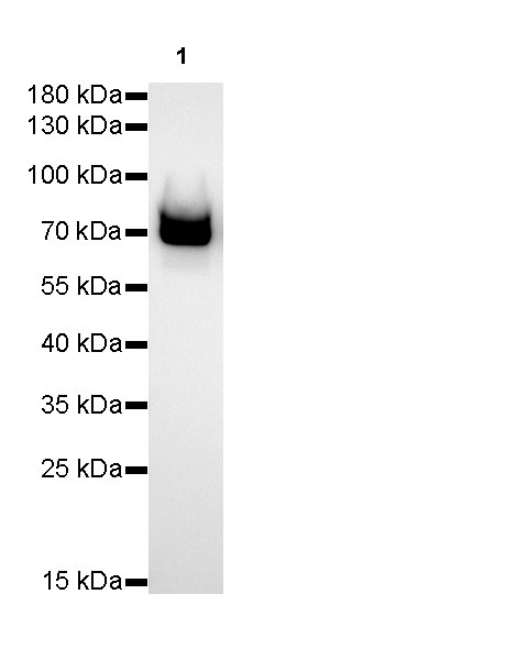
WB result of FUBP1 Rabbit mAb
Primary antibody: FUBP1 Rabbit mAb at 1/5000 dilution
Lane 1: NIH/3T3 whole cell lysate 50 µg
Secondary antibody: Goat Anti-Rabbit IgG, (H+L), HRP conjugated at 1/10000 dilution
Predicted MW: 67 kDa
Observed MW: 70kDa
Exposure time: 180s
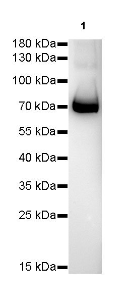
WB result of FUBP1 Rabbit mAb
Primary antibody: FUBP1 Rabbit mAb at 1/5000 dilution
Lane 1: C6 whole cell lysate 50 µg
Secondary antibody: Goat Anti-Rabbit IgG, (H+L), HRP conjugated at 1/10000 dilution
Predicted MW: 67 kDa
Observed MW: 70kDa
Exposure time: 180s
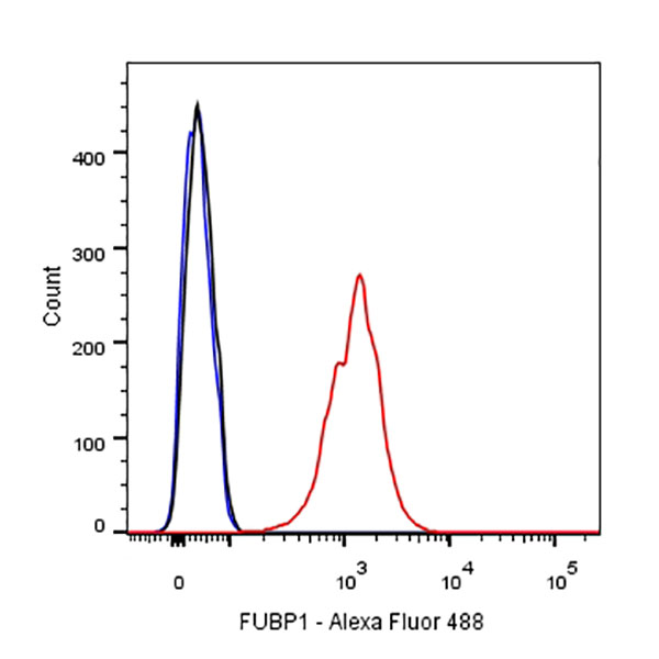
Flow cytometric analysis of HeLa cells labelling FUBP1 antibody at 1/500 (0.1 μg) dilution/ (red) compared with a Rabbit monoclonal IgG (Black) isotype control and an unlabelled control (cells without incubation with primary antibody and secondary antibody) (Blue). Goat Anti-Rabbit IgG Alexa Fluor® 488 was used as the secondary antibody.
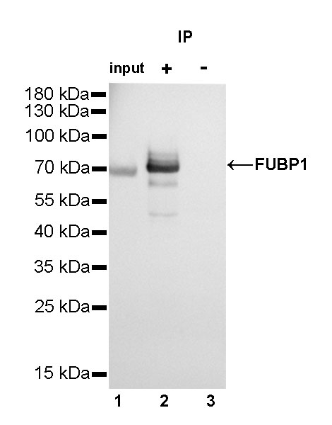
FUBP1 Rabbit mAb at 1/25 dilution (2µg) immunoprecipitating FUBP1 in 0.4mg HeLa whole cell lysate.
Western blot was performed on the immunoprecipitate using FUBP1 Rabbit mAb at 1/1000 dilution.
Secondary antibody (HRP) for IP was used at 1/400 dilution.
Lane 1 : HeLa whole cell lysate 10µg (input)
Lane 2 : FUBP1 Rabbit mAb IP in HeLa whole cell lysate
Lane 3 : Rabbit monoclonal IgG IP in HeLa whole cell lysate
Predicted MW: 67 kDa
Observed MW: 67 kDa
Exposure time: 5s
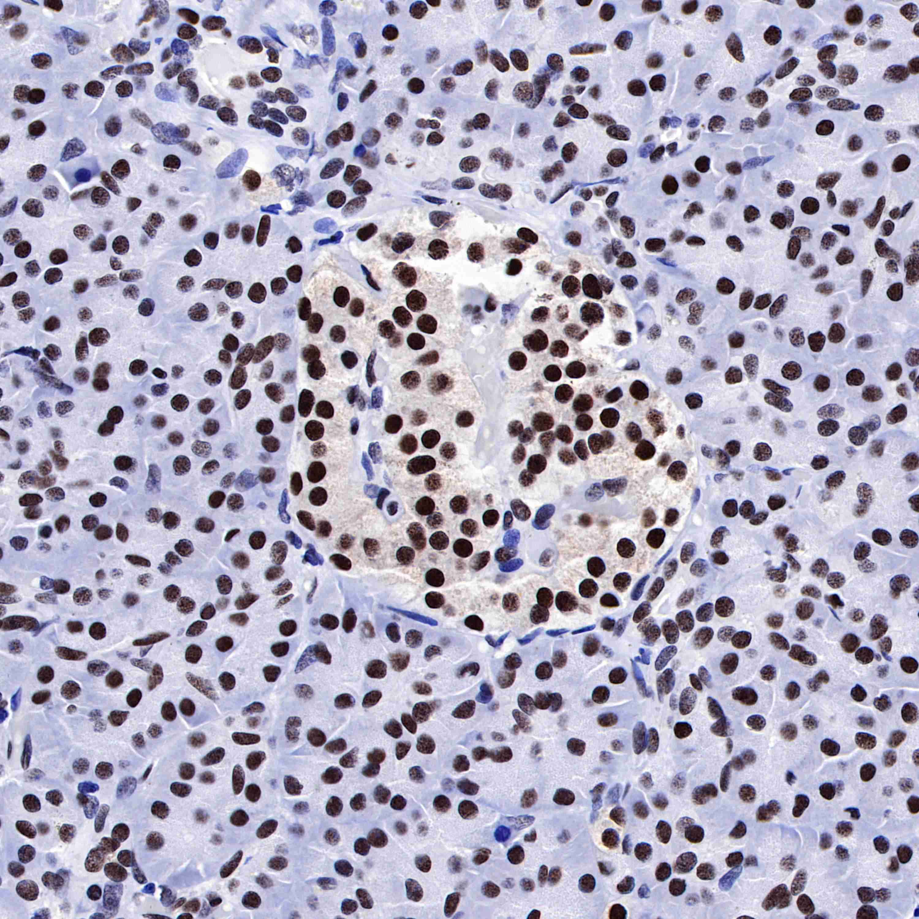
IHC shows positive staining in paraffin-embedded human pancreas. Anti-FUBP1 antibody was used at 1/1000 dilution, followed by a HRP Polymer for Mouse & Rabbit IgG (ready to use). Counterstained with hematoxylin. Heat mediated antigen retrieval with Tris/EDTA buffer pH9.0 was performed before commencing with IHC staining protocol.
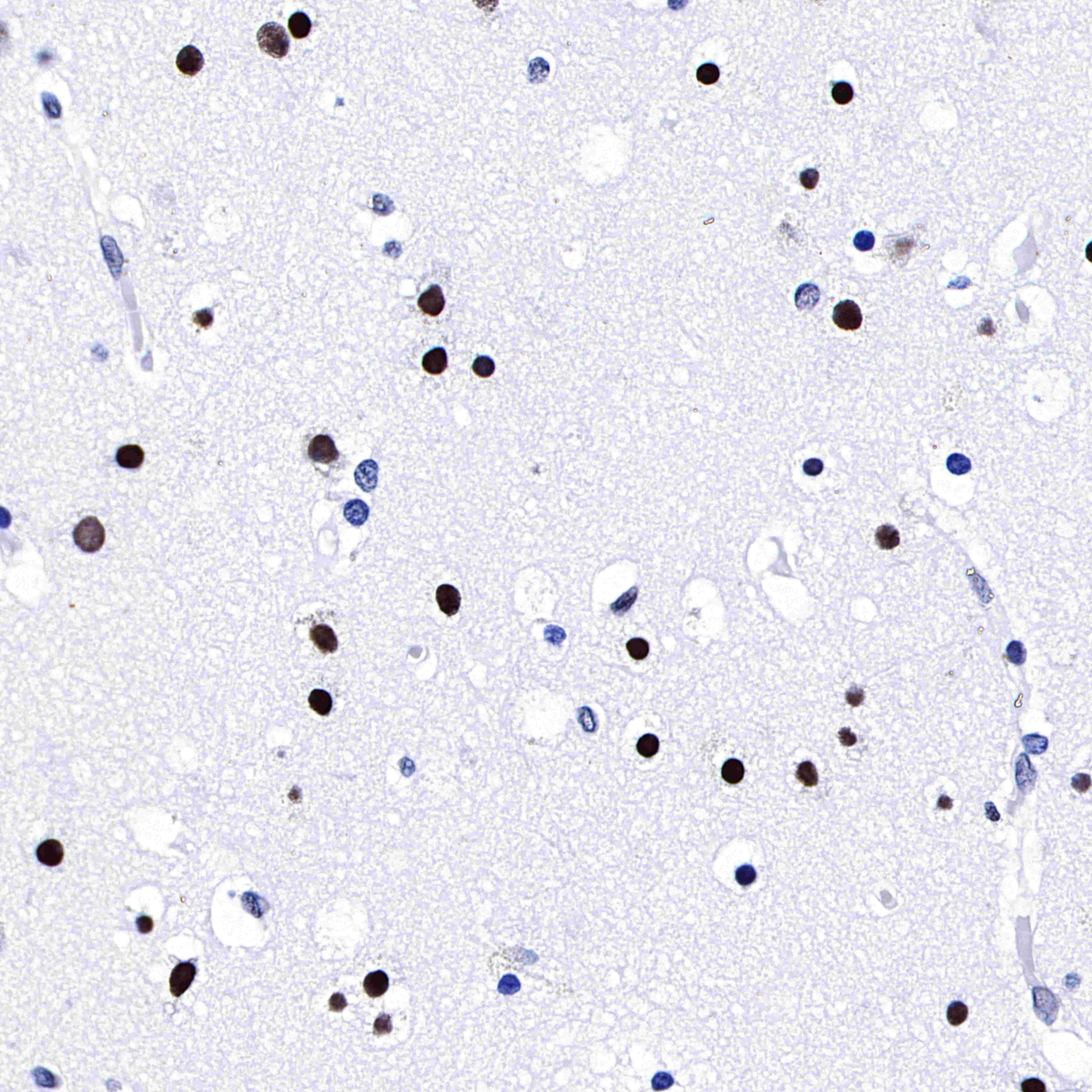
IHC shows positive staining in paraffin-embedded human cerebral cortex. Anti-FUBP1 antibody was used at 1/1000 dilution, followed by a HRP Polymer for Mouse & Rabbit IgG (ready to use). Counterstained with hematoxylin. Heat mediated antigen retrieval with Tris/EDTA buffer pH9.0 was performed before commencing with IHC staining protocol.
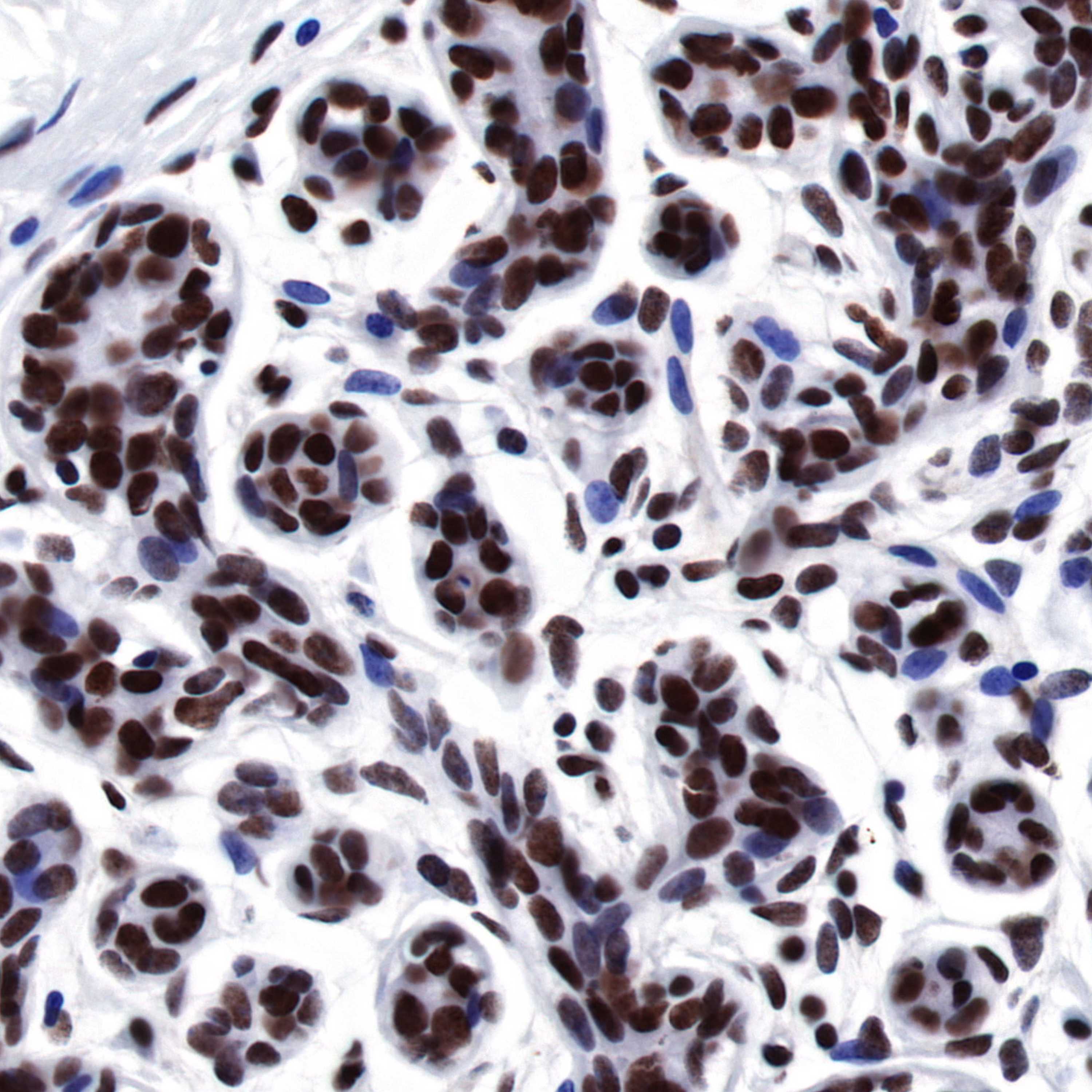
IHC shows positive staining in paraffin-embedded human breast. Anti-FUBP1 antibody was used at 1/1000 dilution, followed by a HRP Polymer for Mouse & Rabbit IgG (ready to use). Counterstained with hematoxylin. Heat mediated antigen retrieval with Tris/EDTA buffer pH9.0 was performed before commencing with IHC staining protocol.
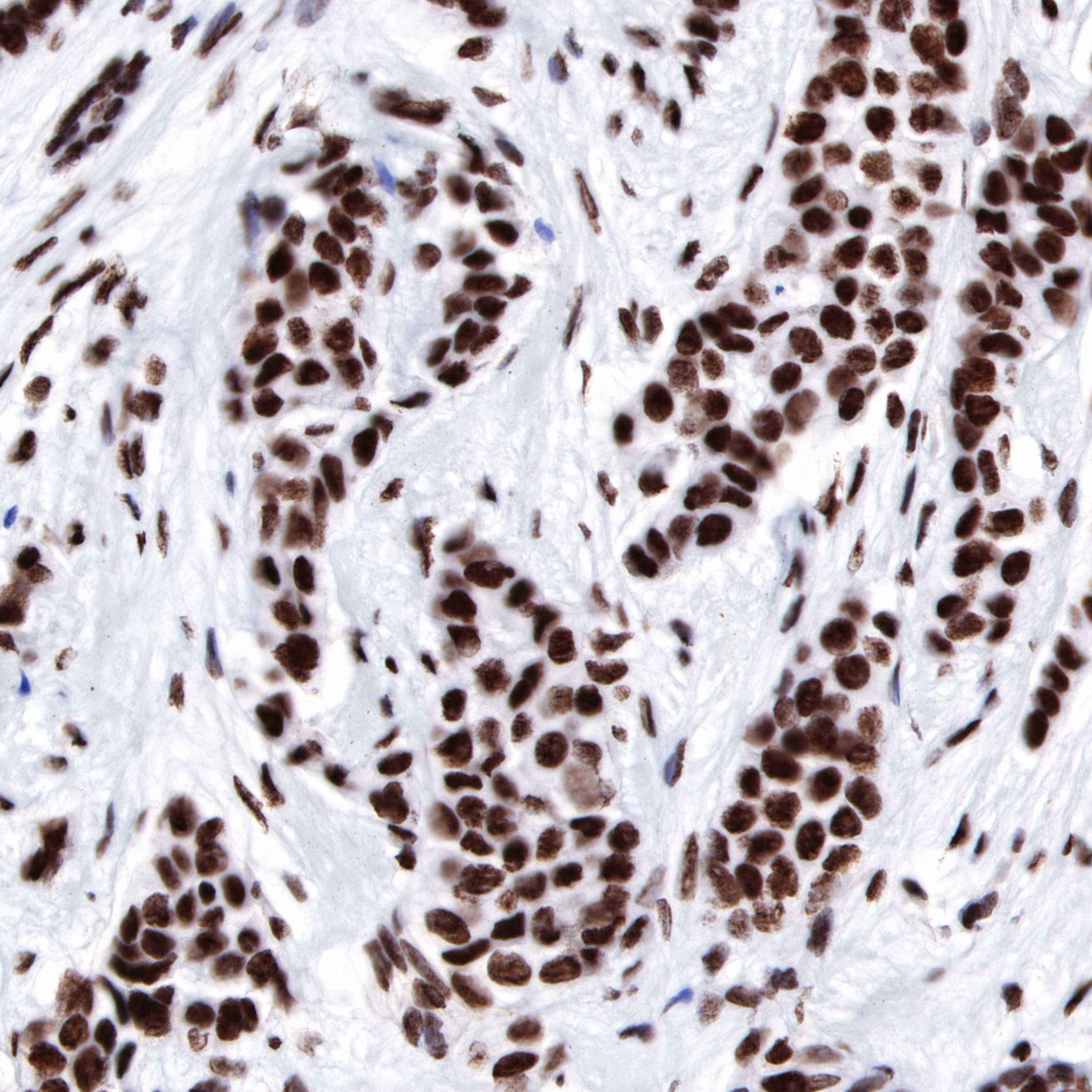
IHC shows positive staining in paraffin-embedded human breast carcinoma. Anti-FUBP1 antibody was used at 1/1000 dilution, followed by a HRP Polymer for Mouse & Rabbit IgG (ready to use). Counterstained with hematoxylin. Heat mediated antigen retrieval with Tris/EDTA buffer pH9.0 was performed before commencing with IHC staining protocol.
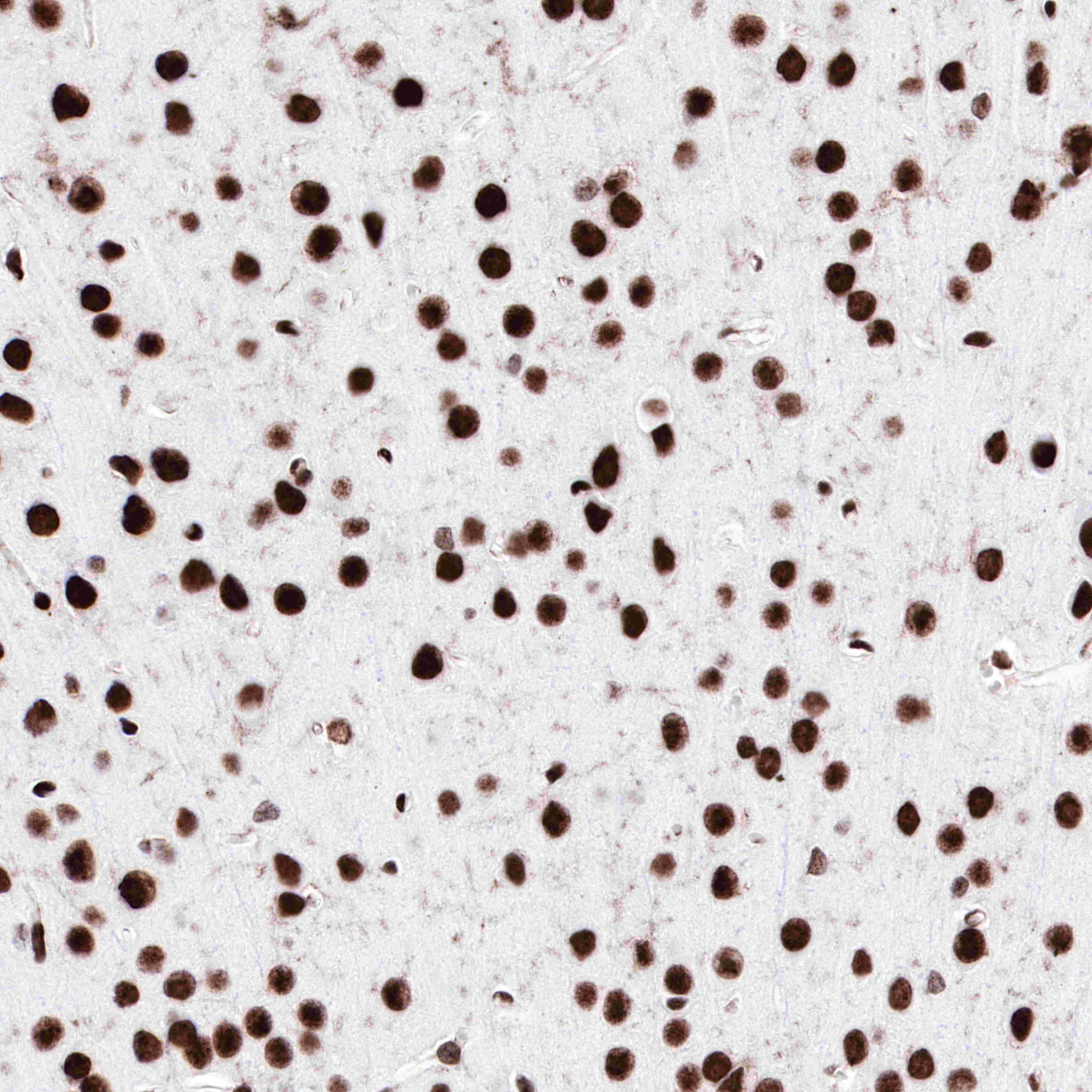
IHC shows positive staining in paraffin-embedded mouse cerebral cortex. Anti-FUBP1 antibody was used at 1/1000 dilution, followed by a HRP Polymer for Mouse & Rabbit IgG (ready to use). Counterstained with hematoxylin. Heat mediated antigen retrieval with Tris/EDTA buffer pH9.0 was performed before commencing with IHC staining protocol.
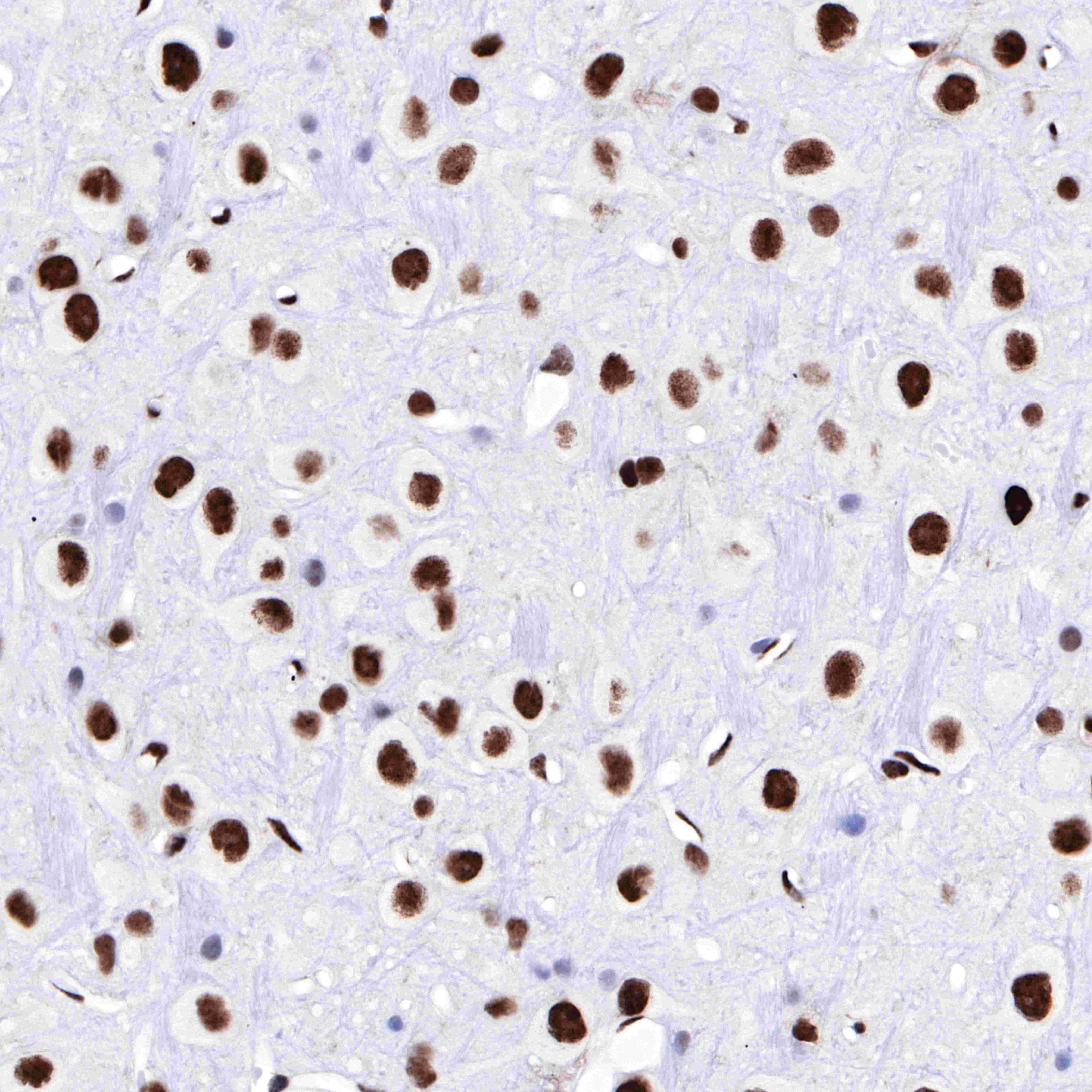
IHC shows positive staining in paraffin-embedded rat cerebral cortex. Anti-FUBP1 antibody was used at 1/1000 dilution, followed by a HRP Polymer for Mouse & Rabbit IgG (ready to use). Counterstained with hematoxylin. Heat mediated antigen retrieval with Tris/EDTA buffer pH9.0 was performed before commencing with IHC staining protocol.
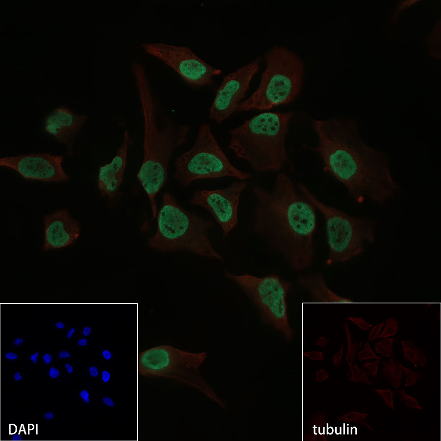
ICC shows positive nuclear staining in HeLa cells. Anti-FUBP1 antibody was used at 1/500 dilution and incubated overnight at 4°C. Goat polyclonal Antibody to Rabbit IgG - H&L (Alexa Fluor® 488) was used as secondary antibody at 1/1000 dilution.The cells were fixed with 4% PFA and permeabilized with 0.1% PBS-Triton X-100. Nuclei were counterstained with DAPI. Counter stain with tubulin (red).
 您现在的位置: