

12 months from date of receipt / reconstitution, -20 °C as supplied
| 应用 | 稀释度 |
|---|---|
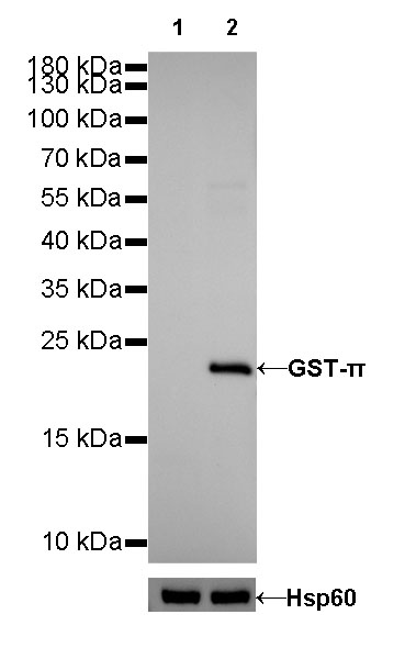
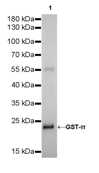
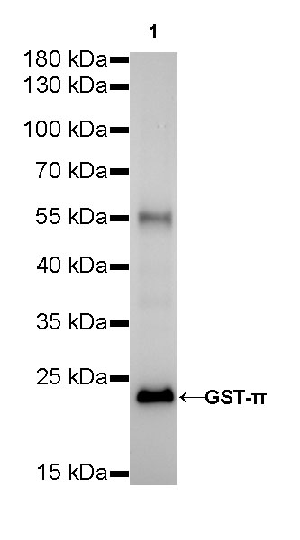
| WB | 1:500-1:2500 |
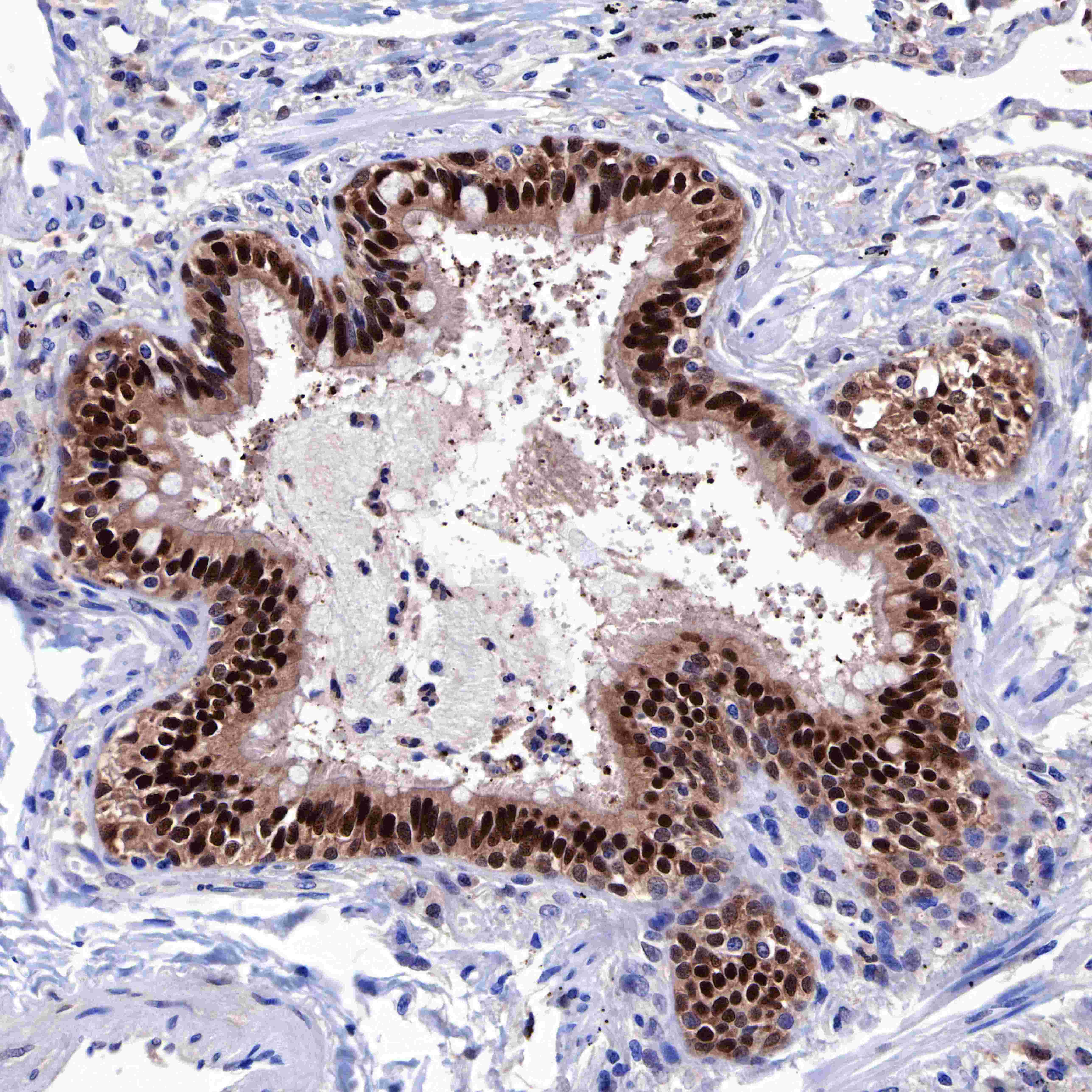
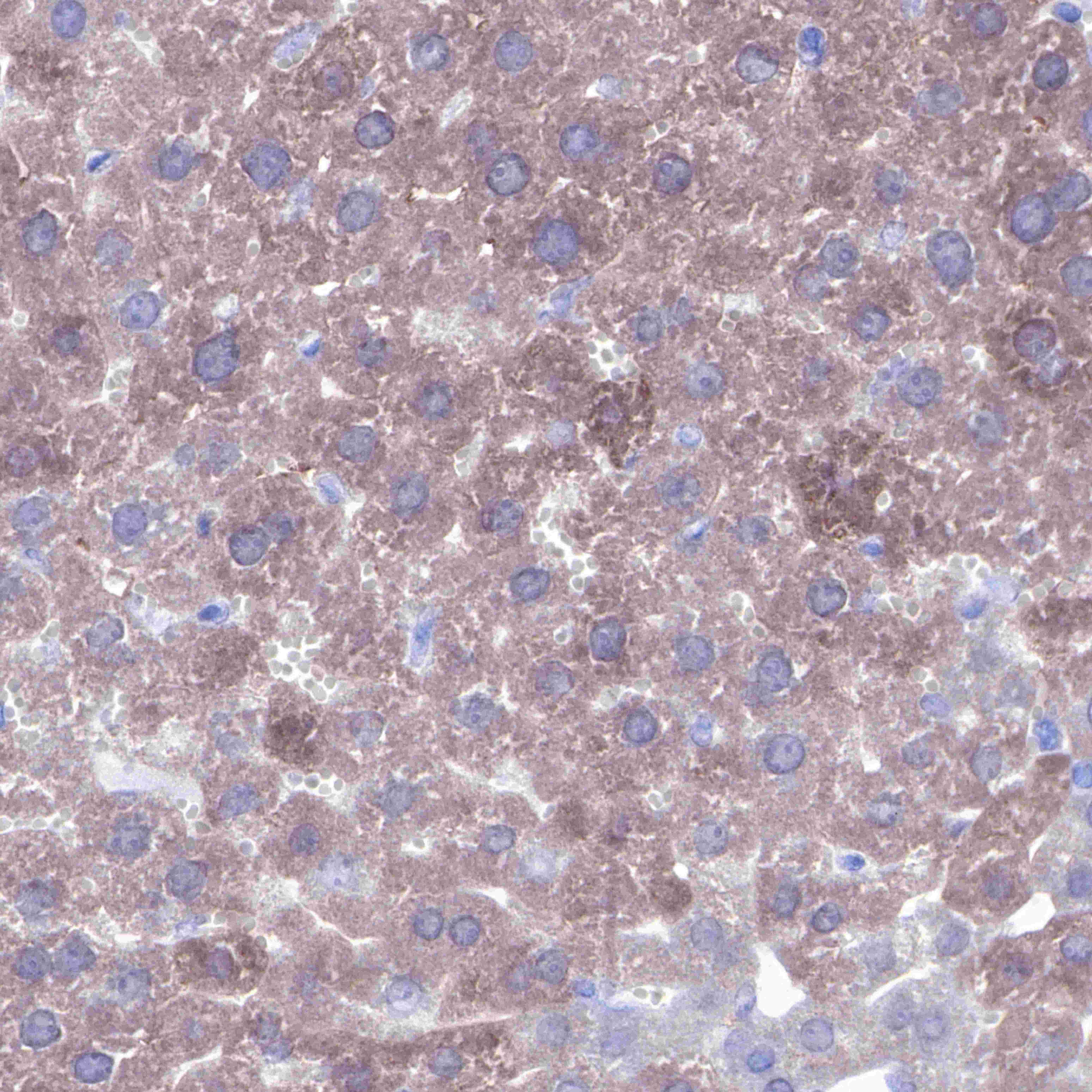
| IHC-P | 1:2000 |
| ICC | 1:50 |
GST-π is abundantly expressed in some mammalian tissues, particularly those associated with malignancies. While the enzyme can catalyze thioether bond formation between some electrophilic chemicals and GSH, novel non-detoxification functions are now ascribed to it.


ICC shows positive staining in K562 cells. Anti-GST-π antibody was used at 1/50 dilution (Green) and incubated overnight at 4°C. Goat polyclonal Antibody to Rabbit IgG - H&L (Alexa Fluor® 488) was used as secondary antibody at 1/1000 dilution. The cells were fixed with 4% PFA and permeabilized with 0.1% PBS-Triton X-100. Nuclei were counterstained with DAPI (Blue). Counterstain with tubulin (red).
 您现在的位置: