

12 months from date of receipt / reconstitution, -20 °C as supplied
| 应用 | 稀释度 |
|---|---|
| ICC | 1:50 |
| IHC-P | 1:1000 |
| WB | 1:500-10000 |
| IP | 1:25 |
| IF | 1:500 |
Carcinoembryonic antigen (CEA) describes a set of highly related glycoproteins involved in cell adhesion. CEA is normally produced in gastrointestinal tissue during fetal development, but the production stops before birth. CEA are glycosyl phosphatidyl inositol (GPI) cell-surface-anchored glycoproteins whose specialized sialofucosylated glycoforms serve as functional colon carcinoma L-selectin and E-selectin ligands, which may be critical to the metastatic dissemination of colon carcinoma cells. Immunologically they are characterized as members of the CD66 cluster of differentiation. The proteins include CD66a, CD66b, CD66c, CD66d, CD66e, CD66f.Consequently, CEA is usually present at very low levels in the blood of healthy adults (about 2–4 ng/mL). However, the serum levels are raised in some types of cancer. CEA is an important tumor marker for colorectal and some other carcinomas. The CEA subgroup members are cell membrane associated and show a complex expression pattern in normal and cancerous tissues with notably CEA showing a selective epithelial expression.
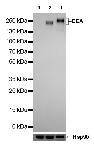
WB result of CEA Rabbit mAb
Primary antibody: CEA Rabbit mAb at 1/500 dilution
Lane 1: PANC-1 whole cell lysate 20 µg
Lane 2: MCF7 whole cell lysate 20 µg
Lane 3: HT-29 whole cell lysate 20 µg
Negative control: PANC-1 whole cell lysate
Secondary antibody: Goat Anti-Rabbit IgG, (H+L), HRP conjugated at 1/10000 dilution
Predicted MW: 200~250 kDa
Observed MW: 200~250 kDa
Exposure time: 180s
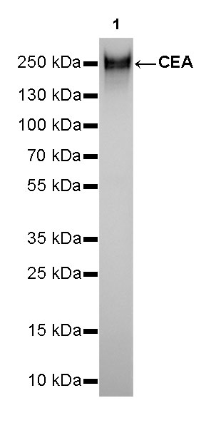
WB result of CEA Rabbit mAb
Primary antibody: CEA Rabbit mAb at 1/10000 dilution
Lane 1: BxPC-3 whole cell lysate 20 µg
Secondary antibody: Goat Anti-Rabbit IgG, (H+L), HRP conjugated at 1/10000 dilution
Predicted MW: 200~250 kDa
Observed MW: 250 kDa
Exposure time: 30s
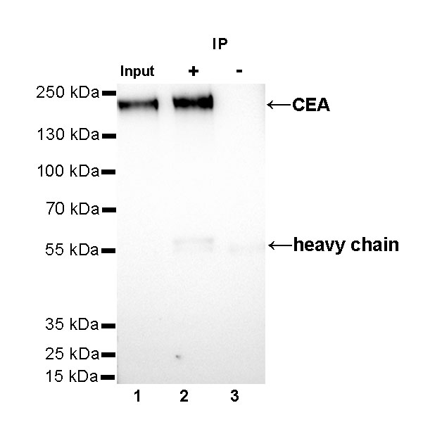
CEA Rabbit mAb at 1/25 dilution (1µg) immunoprecipitating CEA in 0.4mg MCF7 whole cell lysate.
Western blot was performed on the immunoprecipitate using CEA Rabbit mAb at 1/1000 dilution.
Secondary antibody (HRP) for IP was used at 1/400 dilution.
Lane 1 : MCF7 whole cell lysate 30µg(input)
Lane 2 : CEA Rabbit mAb IP in MCF7 whole cell lysate
Lane 3 : Rabbit monoclonal IgG IP in MCF7 whole cell lysate
Predicted MW: 200~250 kDa
Observed MW: 230 kDa
Exposure time: 60s
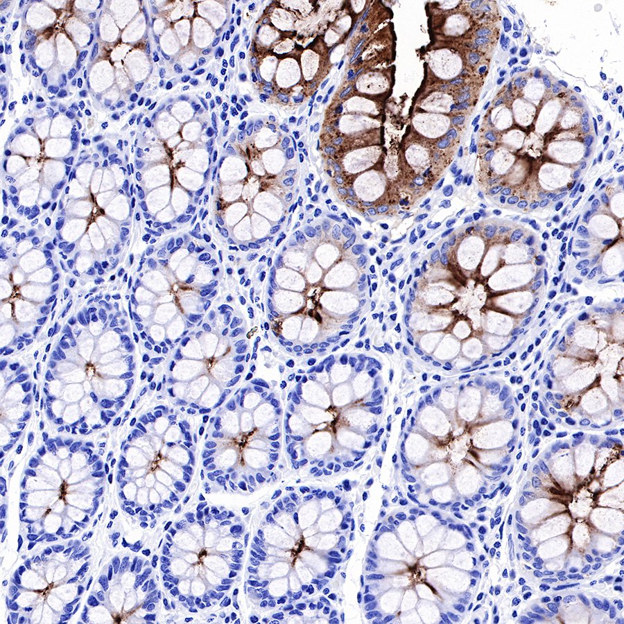
IHC shows positive staining in paraffin-embedded human colon. Anti-CEA(CD66e) antibody was used at 1/1000 dilution, followed by a Goat Anti-Rabbit IgG H&L (HRP) ready to use. Counterstained with hematoxylin. Heat mediated antigen retrieval with Tris/EDTA buffer pH9.0 was performed before commencing with IHC staining protocol.
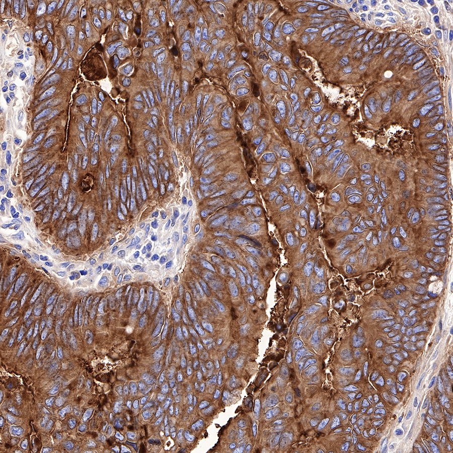
IHC shows positive staining in paraffin-embedded human colon cancer. Anti-CEA(CD66e) antibody was used at 1/1000 dilution, followed by a Goat Anti-Rabbit IgG H&L (HRP) ready to use. Counterstained with hematoxylin. Heat mediated antigen retrieval with Tris/EDTA buffer pH9.0 was performed before commencing with IHC staining protocol.
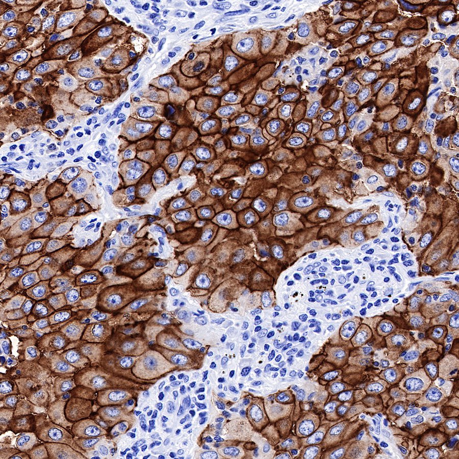
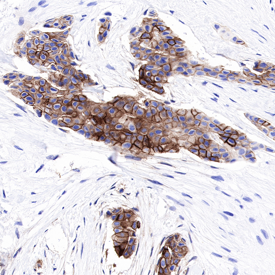
IHC shows positive staining in paraffin-embedded human lung adenocarcinoma. Anti-CEA(CD66e) antibody was used at 1/1000 dilution, followed by a Goat Anti-Rabbit IgG H&L (HRP) ready to use. Counterstained with hematoxylin. Heat mediated antigen retrieval with Tris/EDTA buffer pH9.0 was performed before commencing with IHC staining protocol.
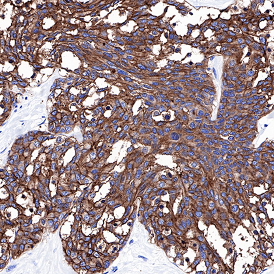
IHC shows positive staining in paraffin-embedded human medullary thyroid carcinoma. Anti-CEA(CD66e) antibody was used at 1/1000 dilution, followed by a Goat Anti-Rabbit IgG H&L (HRP) ready to use. Counterstained with hematoxylin. Heat mediated antigen retrieval with Tris/EDTA buffer pH9.0 was performed before commencing with IHC staining protocol.
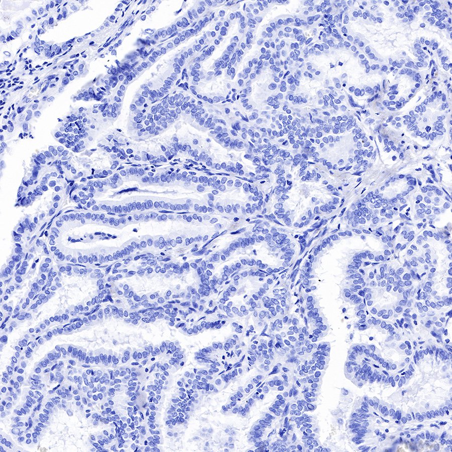
IHC shows negative staining in paraffin-embedded human papillary thyroid carcinoma. Anti-CEA(CD66e) antibody was used at 1/1000 dilution, followed by a Goat Anti-Rabbit IgG H&L (HRP) ready to use. Counterstained with hematoxylin. Heat mediated antigen retrieval with Tris/EDTA buffer pH9.0 was performed before commencing with IHC staining protocol.
IHC shows positive staining in paraffin-embedded human breast cancer. Anti-CEA(CD66e) antibody was used at 1/1000 dilution, followed by a Goat Anti-Rabbit IgG H&L (HRP) ready to use. Counterstained with hematoxylin. Heat mediated antigen retrieval with Tris/EDTA buffer pH9.0 was performed before commencing with IHC staining protocol.
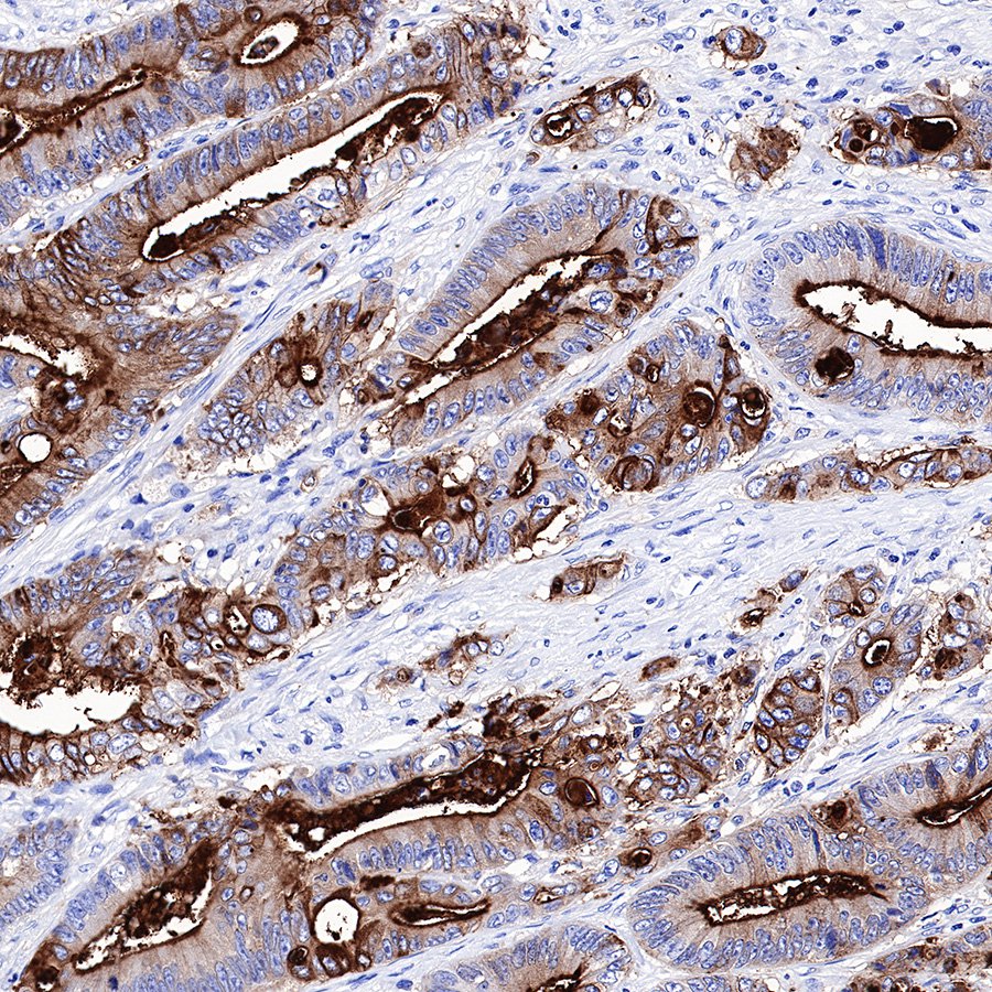
IHC shows positive staining in paraffin-embedded human colon cancer. Anti-CEA(CD66e) antibody was used at 1/1000 dilution, followed by a Goat Anti-Rabbit IgG H&L (HRP) ready to use. Counterstained with hematoxylin. Heat mediated antigen retrieval with Tris/EDTA buffer pH9.0 was performed before commencing with IHC staining protocol.
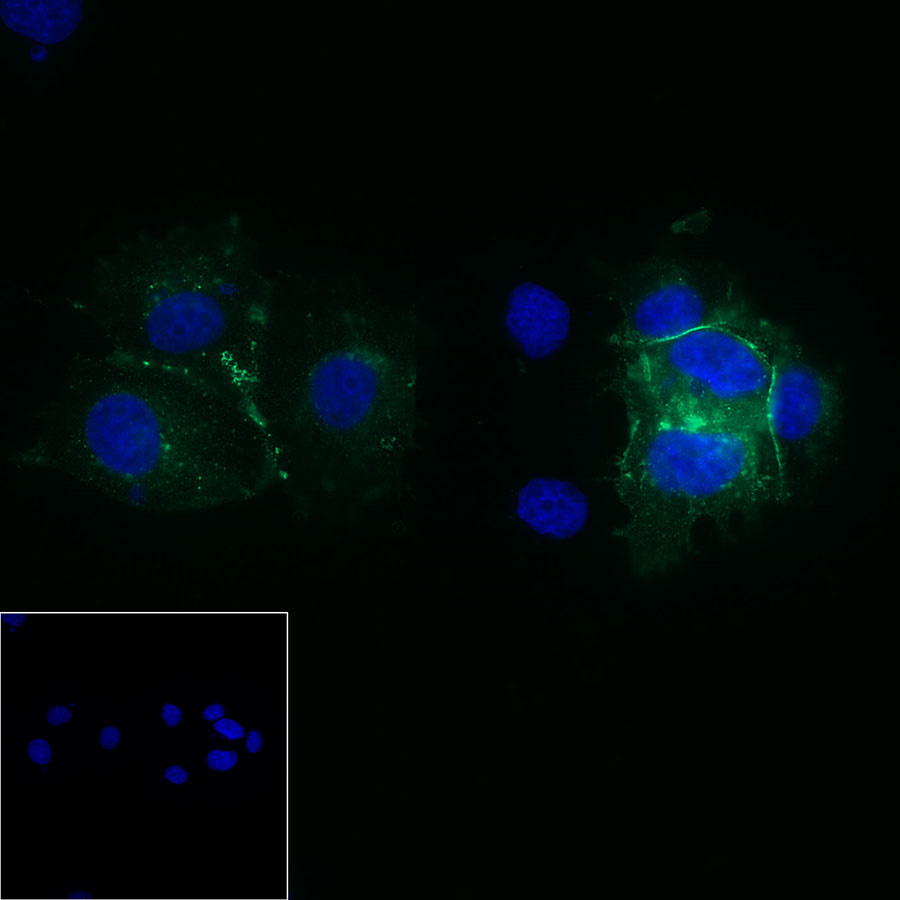
ICC shows positive cell membrane staining in MCF7 cells. Anti-CEA(CD66e) antibody was used at 1/50 dilution and incubated overnight at 4°C. Goat polyclonal Antibody to Rabbit IgG - H&L (Alexa Fluor® 488) was used as secondary antibody at 1/1000 dilution.The cells were fixed with 4% PFA and permeabilized with 0.1% PBS-Triton X-100. Nuclei were counterstained with DAPI.
Negative control: ICC shows negative staining in PANC-1 cells. Anti-CEA(CD66e) antibody was used at 1/50 dilution and incubated overnight at 4°C. Goat polyclonal Antibody to Rabbit IgG - H&L (Alexa Fluor® 488) was used as secondary antibody at 1/1000 dilution.The cells were fixed with 4% PFA and permeabilized with 0.1% PBS-Triton X-100. Nuclei were counterstained with DAPI.
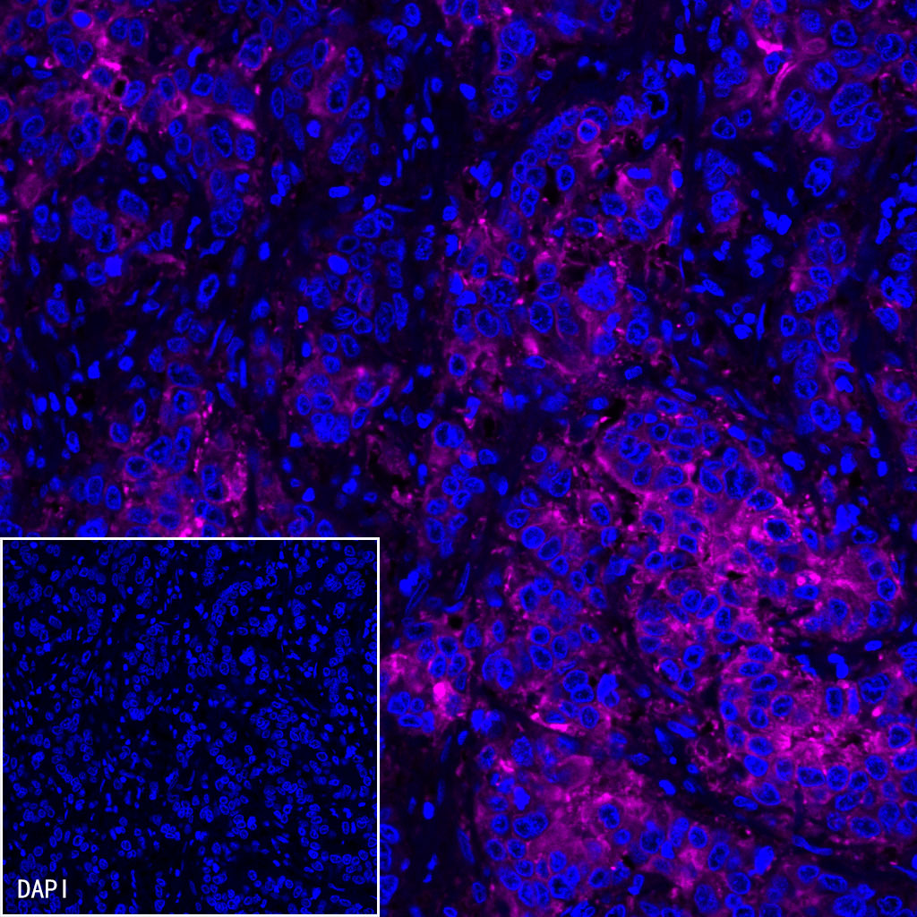
IF shows positive staining in paraffin-embedded human gastric carcinoma. Anti-CEA(CD66e) antibody was used at 1/500 dilution (magenta) and incubated overnight at 4°C. Goat polyclonal Antibody to Rabbit IgG - H&L (Alexa Fluor® 647) was used as secondary antibody at 1/1000 dilution. Counterstained with DAPI (Blue). Heat mediated antigen retrieval with EDTA buffer pH9.0 was performed before commencing with IF staining protocol.

IF shows positive staining in paraffin-embedded human cervical squamous cell carcinoma. Anti-CEA(CD66e) antibody was used at 1/500 dilution (magenta) and incubated overnight at 4°C. Goat polyclonal Antibody to Rabbit IgG - H&L (Alexa Fluor® 647) was used as secondary antibody at 1/1000 dilution. Counterstained with DAPI (Blue). Heat mediated antigen retrieval with EDTA buffer pH9.0 was performed before commencing with IF staining protocol.

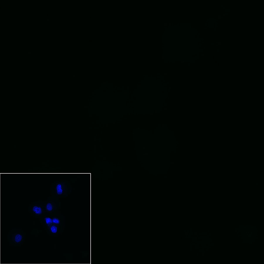
Negative control: IF shows negative staining in paraffin-embedded human papillary thyroid carcinoma. Anti-CEA(CD66e) antibody was used at 1/500 dilution and incubated overnight at 4°C. Goat polyclonal Antibody to Rabbit IgG - H&L (Alexa Fluor® 647) was used as secondary antibody at 1/1000 dilution. Counterstained with DAPI (Blue). Heat mediated antigen retrieval with EDTA buffer pH9.0 was performed before commencing with IF staining protocol.
 您现在的位置: