
| 应用 | 稀释度 |
|---|---|
| WB | 1:1000 |
| ELISA | 5ng/ml-100ng/ml |
Accumulation of intraneuronal neurofibrillary tangles (NFTs) containing paired helical filaments (PHFs) of the microtubule-associated protein tau is one of the defining neuropathological hallmarks of Alzheimer’s disease (AD). The tau protein has an N-terminal projection domain, a proline-rich region, a repeat region, and a C-terminal domain, with multiple potential phosphorylation sites along all regions. Studies using preparations of PHFs and immunohistochemical staining of postmortem brain tissue with specific tau antibodies established that PHF tau is hyperphosphorylated. High levels of p-tau and total tau (t-tau) have consistently been found in cerebrospinal fluid (CSF) of AD patients5. However, while CSF t-tau is considered a non-specific biomarker of neuronal injury, p-tau may reflect AD-related tau pathology in the brain. The vast majority of CSF studies have used immunoassays detecting tau phosphorylated at threonine (Thr) 181 (p-tau181). During the last 2 decades, CSF p-tau181 together with total tau (t-tau) and amyloid-β 42 (Aβ42) have been extensively validated as biomarkers of AD and are currently widely used as diagnostic criteria in research studies, in clinical practice in some countries, and for patient selection in clinical trials. CSF p-tau181 (alone or in combination with Aβ42) accurately differentiates AD from controls and predicts cognitive decline in preclinical and prodromal disease stages. CSF p-tau181 levels are higher in AD compared with other tauopathies including frontotemporal dementia (FTD), progressive supranuclear palsy (PSP) and corticobasal degeneration (CBD) and, hence, CSF p-tau181 has also been proven useful in differential diagnosis of dementia.

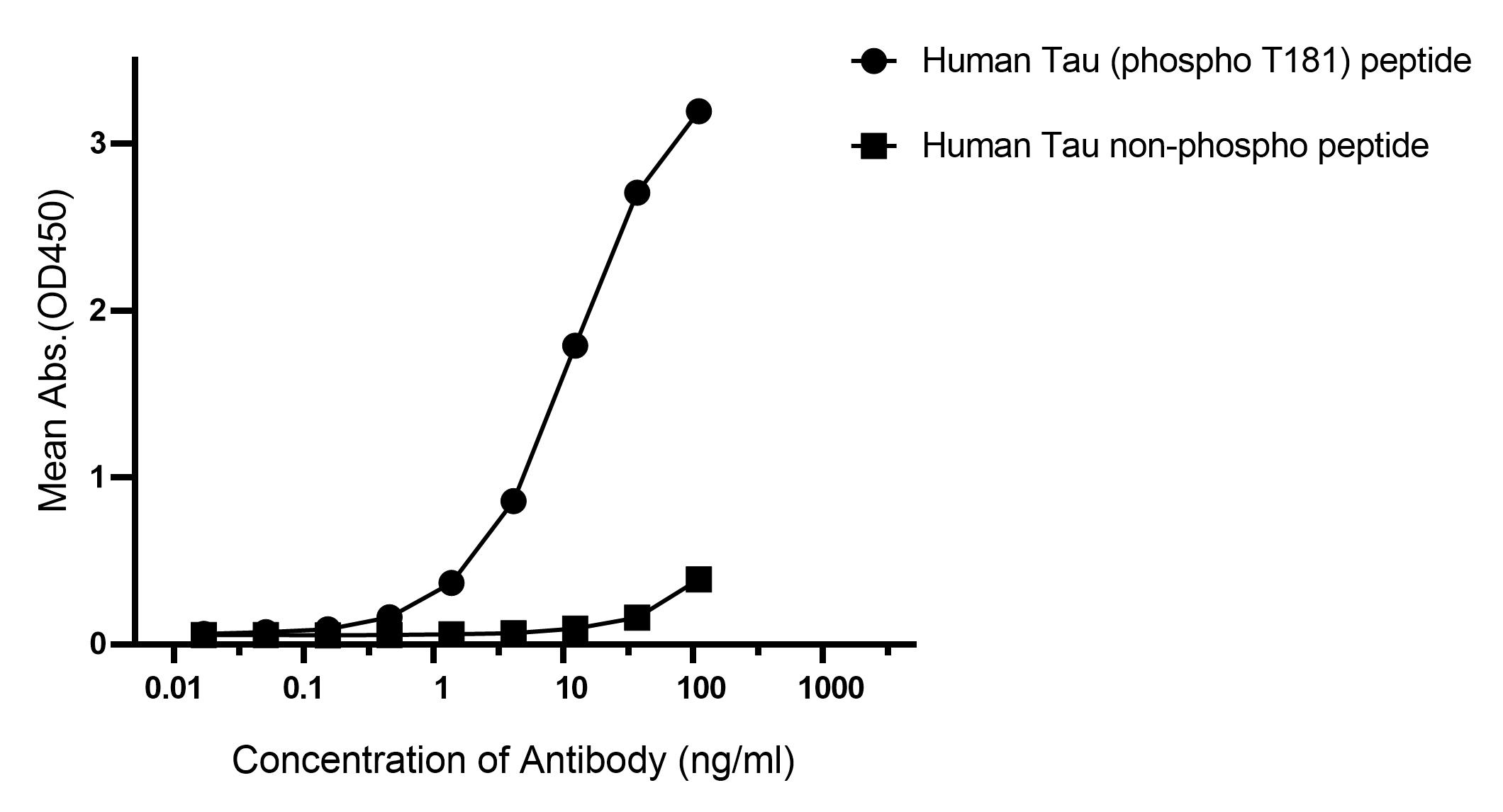
Indirect ELISA antibody dose-response curve using Human Tau non-phospho and Human Tau phospho T181 peptides. Peptide concentration was 500 ng/mL. Tau (phospho T181) Recombinant Rabbit mAb was added at 0-100 ng/mL. Samples were incubated with Peroxidase-AffiniPure Goat Anti-Rabbit IgG (H+L) secondary antibody at 1/10000 dilution.
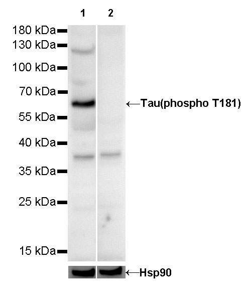
WB result of Tau(phospho T181) Rabbit mAb
Primary antibody: Tau(phospho T181) Rabbit mAb at 1/1000 dilution
Lane 1: old mouse brain lysate 20 µg
Lane 2: old mouse brain lysate(phosphatase treated) 20 µg
Secondary antibody: Goat Anti-Rabbit IgG, (H+L), HRP conjugated at 1/10000 dilution
Predicted MW: 50~70 kDa
Observed MW: 63 kDa
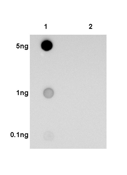
Dot blot result of Tau(phospho T181) Rabbit mAb
Lane 1: Tau phospho T181 peptide
Lane 2: Tau non-phospho T181 peptide
Primary antibody: Tau(phospho T181) Rabbit mAb at 1/1000 dilution
Secondary antibody: Goat Anti-Rabbit IgG, (H+L), HRP conjugated at 1/10000 dilution
Exposure time: 20s
 您现在的位置: