

12 months from date of receipt / reconstitution, -20 °C as supplied
| 应用 | 稀释度 |
|---|---|
| IHC-P | 1:2000 |
| WB | 1:125 |
| ICC | 1:60 |
Alpha-fetoprotein (AFP, α-fetoprotein; also sometimes called alpha-1-fetoprotein, alpha-fetoglobulin, or alpha fetal protein) is a protein that in humans is encoded by the AFP gene. The AFP gene is located on the q arm of chromosome 4 (4q25). Maternal AFP serum level is used to screen for Down syndrome, neural tube defects, and other chromosomal abnormalities. AFP is a major plasma protein produced by the yolk sac and the fetal liver during fetal development. It is thought to be the fetal analog of serum albumin. AFP binds to copper, nickel, fatty acids and bilirubin and is found in monomeric, dimeric and trimeric forms.
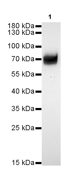
WB result of AFP Rabbit mAb
Primary antibody: AFP Rabbit mAb at 1/125 dilution
Lane 1: mouse placenta lysate 20 µg
Secondary antibody: Goat Anti-Rabbit IgG, (H+L), HRP conjugated at 1/10000 dilution
Predicted MW: 70kDa
Observed MW: 70kDa
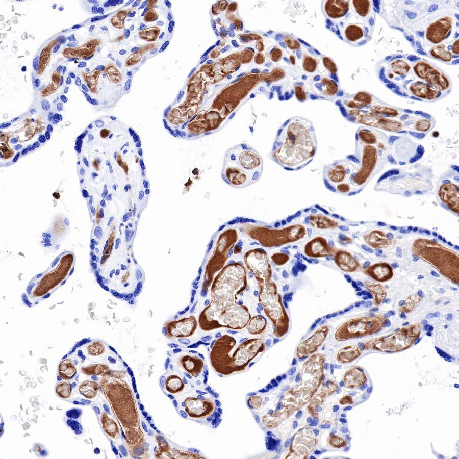
IHC shows positive staining in paraffin-embedded human placenta.
Anti-AFP antibody was used at 1/2000 dilution, followed by a Goat Anti-Rabbit IgG H&L (HRP) ready to use.
Counterstained with hematoxylin.
Heat mediated antigen retrieval with Tris/EDTA buffer pH9.0 was performed before commencing with IHC staining protocol.
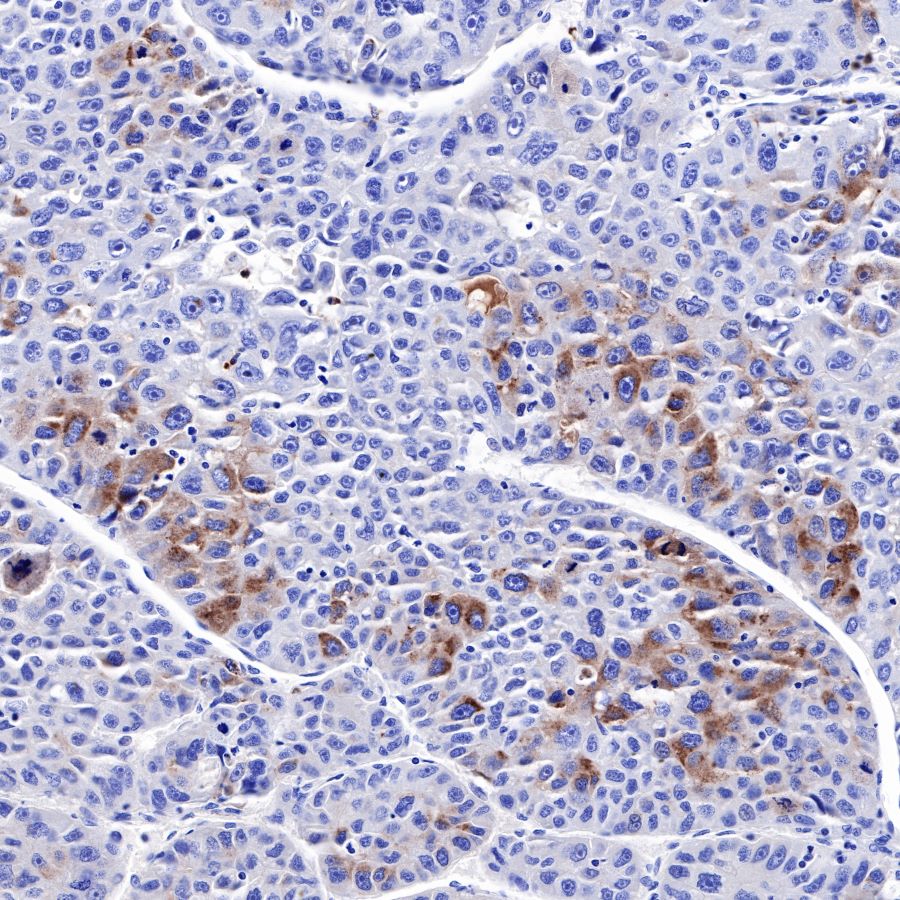
IHC shows positive staining in paraffin-embedded human hepatocellular carcinoma.
Anti-AFP antibody was used at 1/2000 dilution, followed by a Goat Anti-Rabbit IgG H&L (HRP) ready to use.
Counterstained with hematoxylin.
Heat mediated antigen retrieval with Tris/EDTA buffer pH9.0 was performed before commencing with IHC staining protocol.
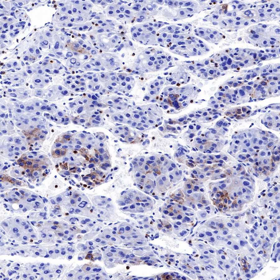
IHC shows positive staining in paraffin-embedded human hepatocellular carcinoma.
Anti-AFP antibody was used at 1/2000 dilution, followed by a Goat Anti-Rabbit IgG H&L (HRP) ready to use.
Counterstained with hematoxylin.
Heat mediated antigen retrieval with Tris/EDTA buffer pH9.0 was performed before commencing with IHC staining protocol.
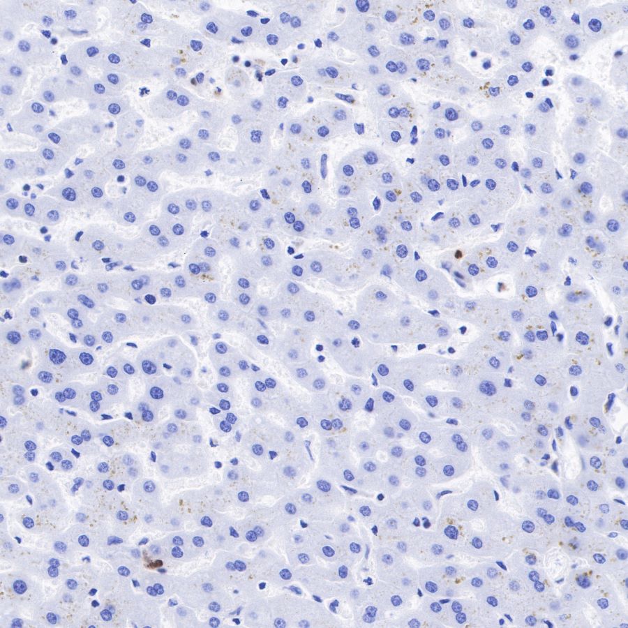
Negative control: IHC shows negative staining in paraffin-embedded human liver.
Anti-AFP antibody was used at 1/2000 dilution, followed by a Goat Anti-Rabbit IgG H&L (HRP) ready to use.
Counterstained with hematoxylin.
Heat mediated antigen retrieval with Tris/EDTA buffer pH9.0 was performed before commencing with IHC staining protocol.
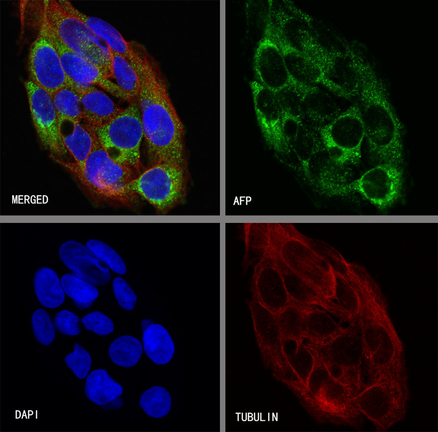
ICC shows positive staining in HepG2 cells. Anti-AFP antibody was used at 1/60 dilution (Green) and incubated overnight at 4°C. Goat polyclonal Antibody to Rabbit IgG - H&L (Alexa Fluor® 488) was used as secondary antibody at 1/1000 dilution. The cells were fixed with 100% ice-cold methanol and permeabilized with 0.1% PBS-Triton X-100. Nuclei were counterstained with DAPI (Blue).Counterstain with tubulin (red).
 您现在的位置: