
12 months from date of receipt / reconstitution, -20 °C as supplied
| 应用 | 稀释度 |
|---|---|
| IP | 1:25 |
| IHC-P | 1:2000 |
| WB | 1:1000 |
| ICFCM | 1:500 |
| ICC | 1:500 |
Heat shock protein HSP 90-beta also called HSP90beta is a protein that in humans is encoded by the HSP90AB1 gene. HSP90AB1 is a molecular chaperone. Chaperones are proteins that bind to other proteins, thereby stabilizing them in an ATP-dependent manner. Chaperones stabilize new proteins during translation, mature proteins which are partially unstable but also proteins that have become partially denatured due to various kinds of cellular stress. In case proper folding or refolding is impossible, HSPs mediate protein degradation. They also have specialized functions, such as intracellular transport into organelles. HSP90AB1 and its co-chaperones are frequently overexpressed in cancer cells. They are able to stabilize mutant proteins thereby allowing survival and increased proliferation of cancer cells. This renders HSPs potential targets for cancer treatment. In salivary gland tumors, expression of HSP90AA1 and HSP90AB1 correlates with malignancy, proliferation and metastasis. The same is basically true for lung cancers where a correlation with survival was found.

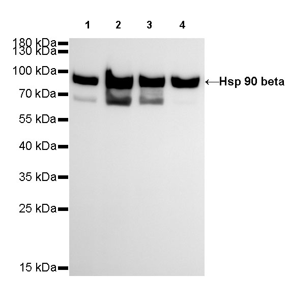
WB result of Hsp90 beta Rabbit mAb
Primary antibody: Hsp90 beta Rabbit mAb at 1/1000 dilution
Lane 1: Hela whole cell lysate 20 µg
Lane 2: K562 whole cell lysate 20 µg
Lane 3: Jurkat whole cell lysate 20 µg
Lane 4: SH-SY5Y whole cell lysate 20 µg
Secondary antibody: Goat Anti-Rabbit IgG, (H+L), HRP conjugated at 1/10000 dilution
Predicted MW: 90 kDa
Observed MW: 90 kDa
Exposure time: 24s
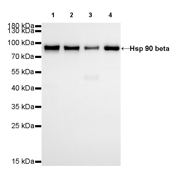
WB result of Hsp90 beta Rabbit mAb
Primary antibody: Hsp90 beta Rabbit mAb at 1/1000 dilution
Lane 1: mouse brain lysate 20 µg
Lane 2: mouse kidney lysate 20 µg
Lane 3: mouse heart lysate 20 µg
Lane 4: mouse spleen lysate 20 µg
Secondary antibody: Goat Anti-Rabbit IgG, (H+L), HRP conjugated at 1/10000 dilution
Predicted MW: 90 kDa
Observed MW: 90 kDa
Exposure time:Lane 1~4:180s
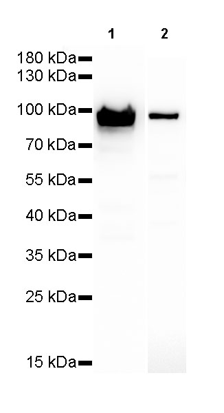
WB result of Hsp90 beta Rabbit mAb
Primary antibody: Hsp90 beta Rabbit mAb at 1/1000 dilution
Lane 1: PC-12 whole cell lysate 20 µg
Lane 2: rat skin lysate 20 µg
Secondary antibody: Goat Anti-Rabbit IgG, (H+L), HRP conjugated at 1/10000 dilution
Predicted MW: 90 kDa
Observed MW: 90 kDa
Exposure time: lane1: 30s; lane1: 180s

Flow cytometric analysis of 4% PFA fixed 90% methanol permeabilized Jurkat (Human T cell leukemia T lymphocyte) labelling Hsp90 beta antibody at 1/500 dilution (0.1 μg) / (Red) compared with a Rabbit monoclonal IgG (Black) isotype control and an unlabelled control (cells without incubation with primary antibody and secondary antibody) (Blue). Goat Anti - Rabbit IgG Alexa Fluor® 488 was used as the secondary antibody.
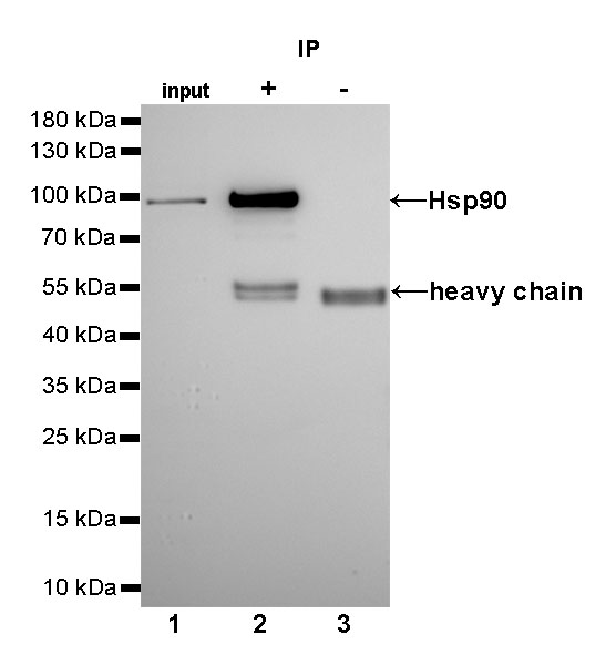
Hsp90 beta Rabbit mAb at 1/25 dilution (2µg) immunoprecipitating Hsp90 beta in 0.4mg HeLa whole cell lysate.
Western blot was performed on the immunoprecipitate using Hsp90 beta Rabbit mAb at 1/1000 dilution.
Secondary antibody (HRP) for IP was used at 1/400 dilution.
Lane 1: HeLa whole cell lysate 10µg (input)
Lane 2 (+): Hsp90 beta Rabbit mAb IP in HeLa whole cell lysate
Lane 3 (-): Rabbit monoclonal IgG IP in HeLa whole cell lysate
Predicted MW: 90 kDa
Observed MW: 90 kDa
Exposure time: 30s
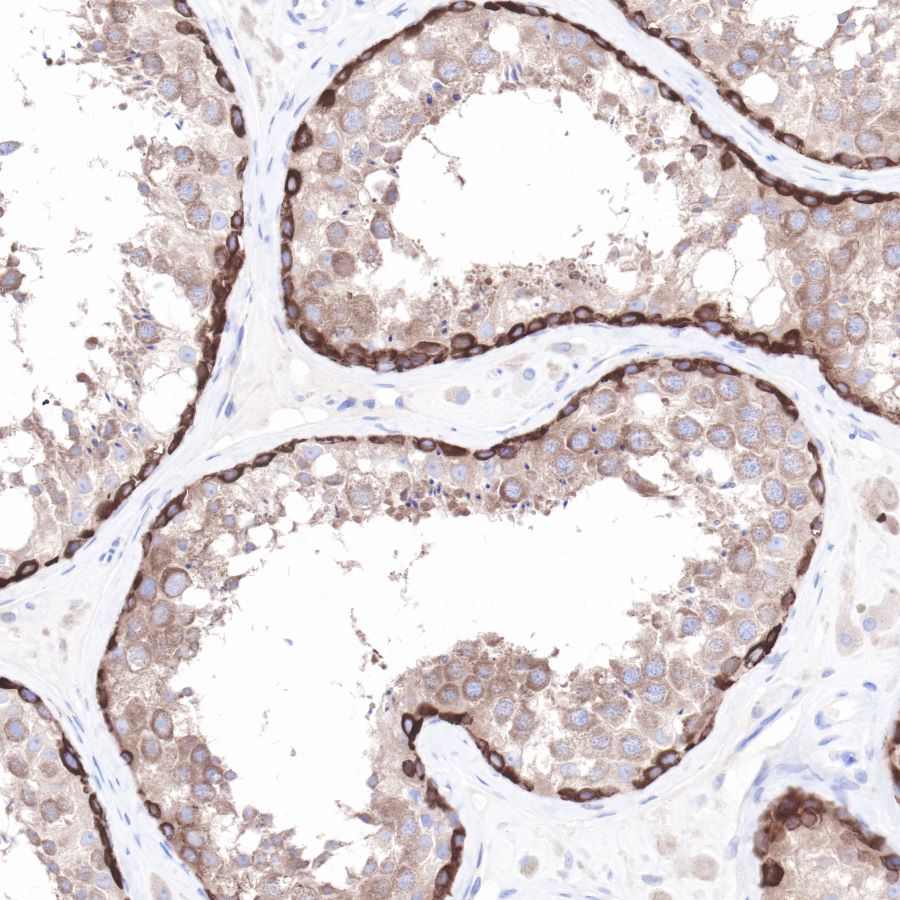
IHC shows positive staining in paraffin-embedded human testis.
Anti-Hsp90 beta antibody was used at 1/2000 dilution, followed by a Goat Anti-Rabbit IgG H&L (HRP) ready to use. Counterstained with hematoxylin.
Heat mediated antigen retrieval with Tris/EDTA buffer pH9.0 was performed before commencing with IHC staining protocol.
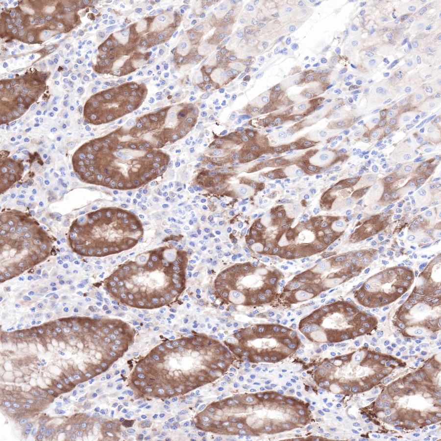
IHC shows positive staining in paraffin-embedded human stomach.
Anti-Hsp90 beta antibody was used at 1/2000 dilution, followed by a Goat Anti-Rabbit IgG H&L (HRP) ready to use. Counterstained with hematoxylin.
Heat mediated antigen retrieval with Tris/EDTA buffer pH9.0 was performed before commencing with IHC staining protocol.
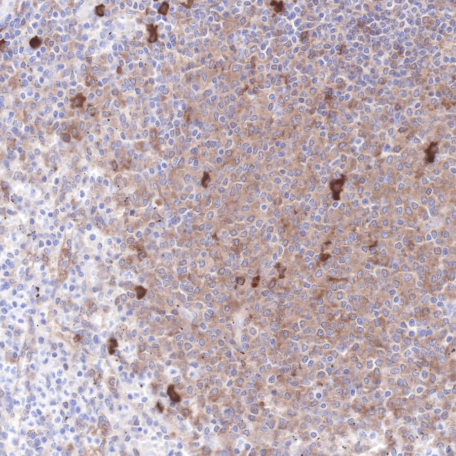
IHC shows positive staining in paraffin-embedded human spleen.
Anti-Hsp90 beta antibody was used at 1/2000 dilution, followed by a Goat Anti-Rabbit IgG H&L (HRP) ready to use. Counterstained with hematoxylin.
Heat mediated antigen retrieval with Tris/EDTA buffer pH9.0 was performed before commencing with IHC staining protocol.
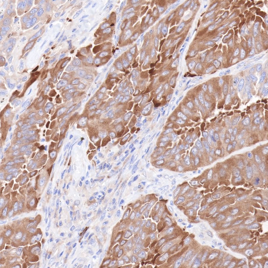
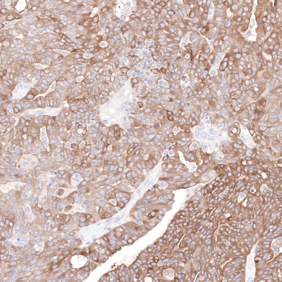
IHC shows positive staining in paraffin-embedded human lung cancer.
Anti-Hsp90 beta antibody was used at 1/2000 dilution, followed by a Goat Anti-Rabbit IgG H&L (HRP) ready to use. Counterstained with hematoxylin.
Heat mediated antigen retrieval with Tris/EDTA buffer pH9.0 was performed before commencing with IHC staining protocol.
IHC shows positive staining in paraffin-embedded human ovarian cancer.
Anti-Hsp90 beta antibody was used at 1/2000 dilution, followed by a Goat Anti-Rabbit IgG H&L (HRP) ready to use. Counterstained with hematoxylin.
Heat mediated antigen retrieval with Tris/EDTA buffer pH9.0 was performed before commencing with IHC staining protocol.
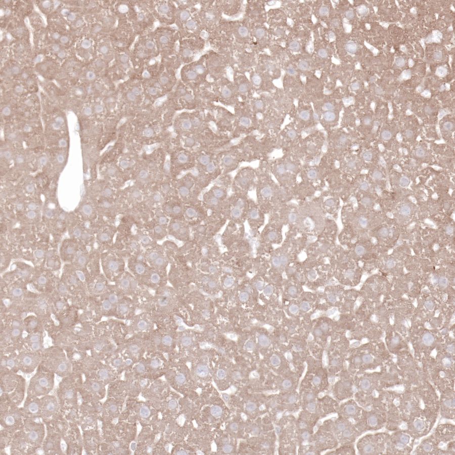
IHC shows positive staining in paraffin-embedded mouse liver.
Anti-Hsp90 beta antibody was used at 1/2000 dilution, followed by a Goat Anti-Rabbit IgG H&L (HRP) ready to use. Counterstained with hematoxylin.
Heat mediated antigen retrieval with Tris/EDTA buffer pH9.0 was performed before commencing with IHC staining protocol.
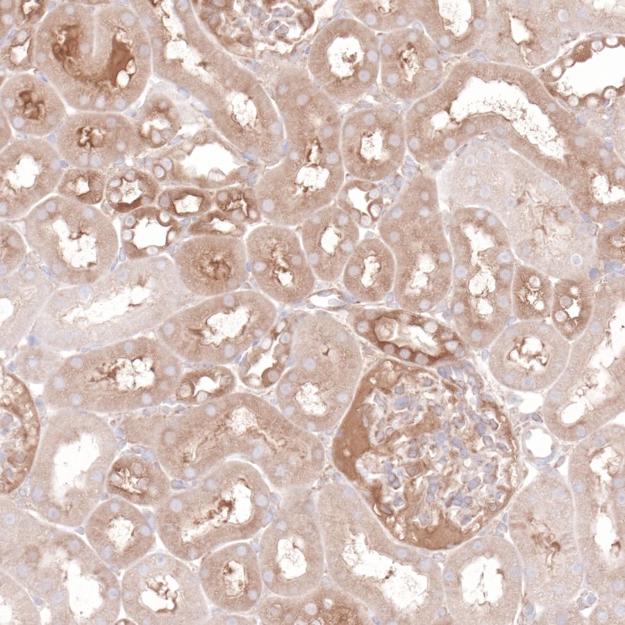
IHC shows positive staining in paraffin-embedded rat kidney.
Anti-Hsp90 beta antibody was used at 1/2000 dilution, followed by a Goat Anti-Rabbit IgG H&L (HRP) ready to use. Counterstained with hematoxylin.
Heat mediated antigen retrieval with Tris/EDTA buffer pH9.0 was performed before commencing with IHC staining protocol.
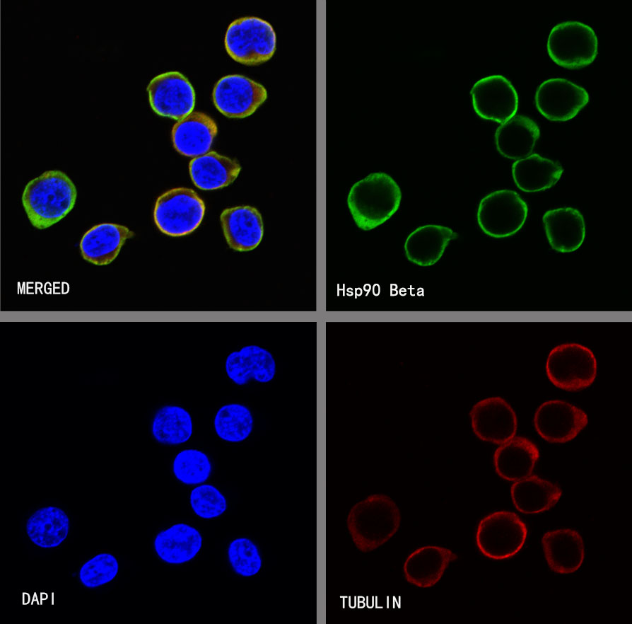
ICC shows positive staining in Jurkat cells. Anti-Hsp90 beta antibody was used at 1/500 dilution (Green) and incubated overnight at 4°C. Goat polyclonal Antibody to Rabbit IgG - H&L (Alexa Fluor® 488) was used as secondary antibody at 1/1000 dilution. The cells were fixed with 4% PFA and permeabilized with 0.1% PBS-Triton X-100. Nuclei were counterstained with DAPI (Blue).Counterstain with tubulin (Red).
 您现在的位置: