
12 months from date of receipt / reconstitution, -20 °C as supplied
| 应用 | 稀释度 |
|---|---|
| WB | 1:1000 |
| IHC-P | 1:500 |
| ICC | 1:200-1:500 |
Homeobox protein CDX-2 is a protein that in humans is encoded by the CDX2 gene. The CDX2 protein is a homeobox transcription factor expressed in the nuclei of intestinal epithelial cells, playing an essential role in the development and function of the digestive system. CDX2 is also used in diagnostic surgical pathology as a marker for gastrointestinal differentiation, especially colorectal.
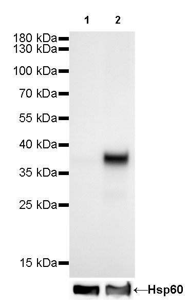
WB result of CDX2 Rabbit mAb
Primary antibody:CDX2 Rabbit mAb at 1/1000 dilution
Lane 1: Hela whole cell lysate 20 µg
Lane 2: Caco2 whole cell lysate 20 µg
Negative control: Hela whole cell lysate
Secondary antibody: Goat Anti-Rabbit IgG, (H+L), HRP conjugated at 1/10000 dilution
Predicted MW: 38 kDa
Observed MW: 38 kDa
Exposure time: 60s
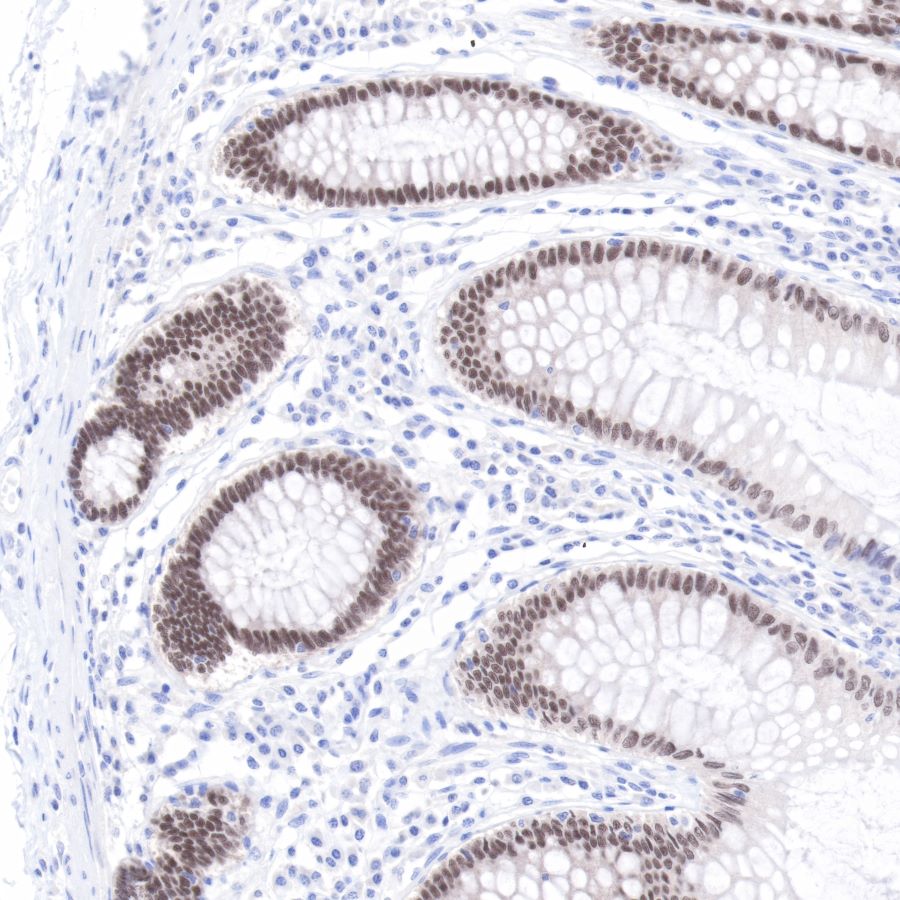
IHC shows positive staining in paraffin-embedded human colon.
Anti-CDX2 antibody was used at 1/500 dilution, followed by a Goat Anti-Rabbit IgG H&L (HRP) ready to use. Counterstained with hematoxylin.
Heat mediated antigen retrieval with Tris/EDTA buffer pH9.0 was performed before commencing with IHC staining protocol.
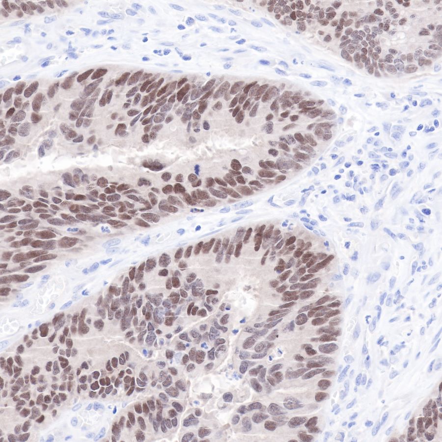
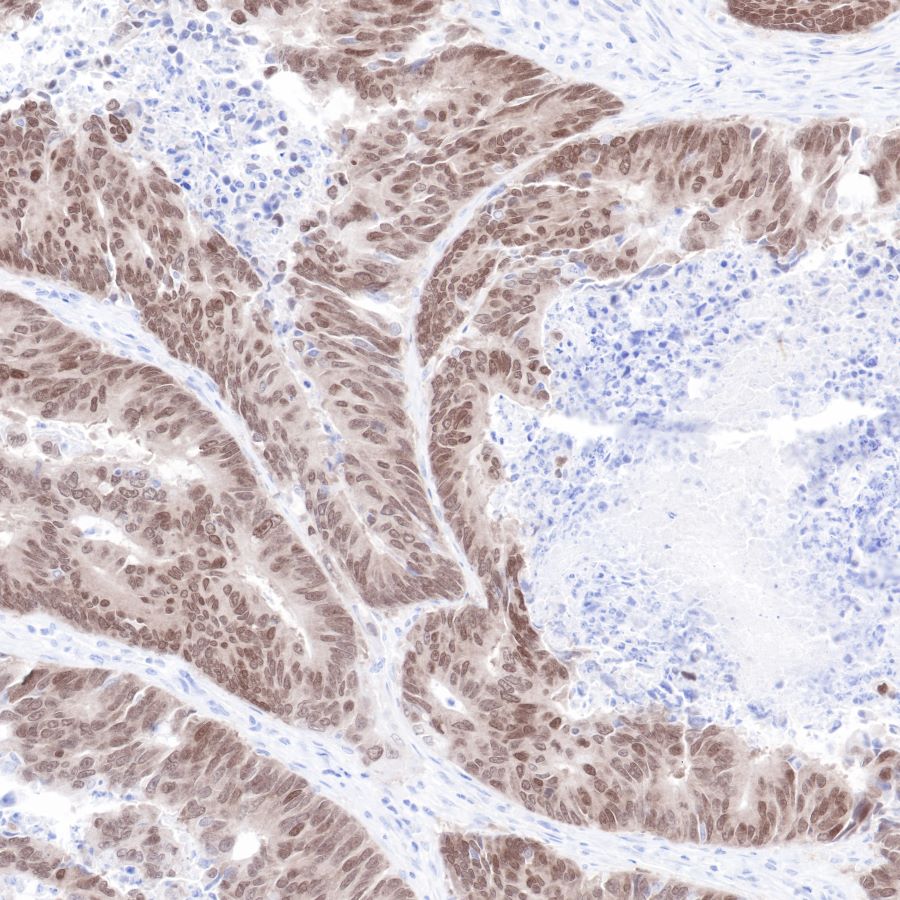
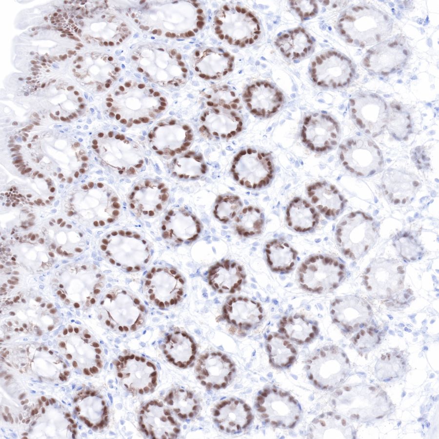
IHC shows positive staining in paraffin-embedded human colon cancer.
Anti-CDX2 antibody was used at 1/500 dilution, followed by a Goat Anti-Rabbit IgG H&L (HRP) ready to use. Counterstained with hematoxylin.
Heat mediated antigen retrieval with Tris/EDTA buffer pH9.0 was performed before commencing with IHC staining protocol.
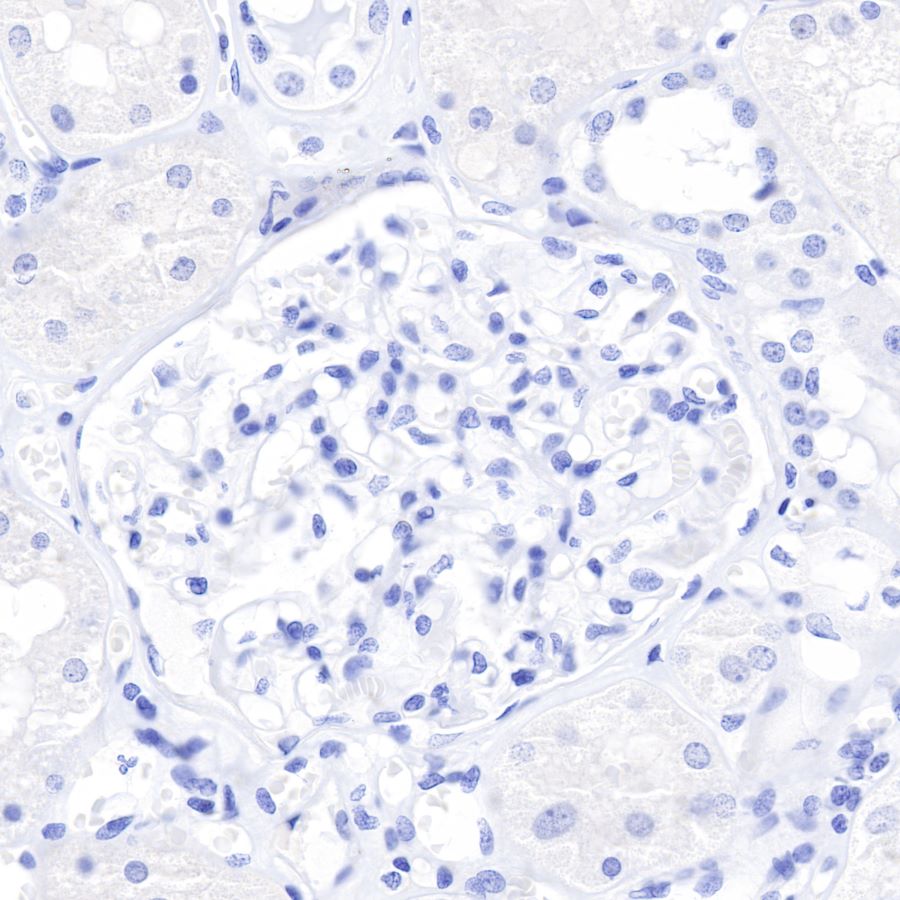
IHC shows negative staining in paraffin-embedded human kidney.
Anti-CDX2 antibody was used at 1/500 dilution, followed by a Goat Anti-Rabbit IgG H&L (HRP) ready to use. Counterstained with hematoxylin.
Heat mediated antigen retrieval with Tris/EDTA buffer pH9.0 was performed before commencing with IHC staining protocol.
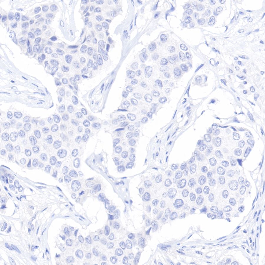
IHC shows negative staining in paraffin-embedded human breast cancer.
Anti-CDX2 antibody was used at 1/500 dilution, followed by a Goat Anti-Rabbit IgG H&L (HRP) ready to use. Counterstained with hematoxylin.
Heat mediated antigen retrieval with Tris/EDTA buffer pH9.0 was performed before commencing with IHC staining protocol.
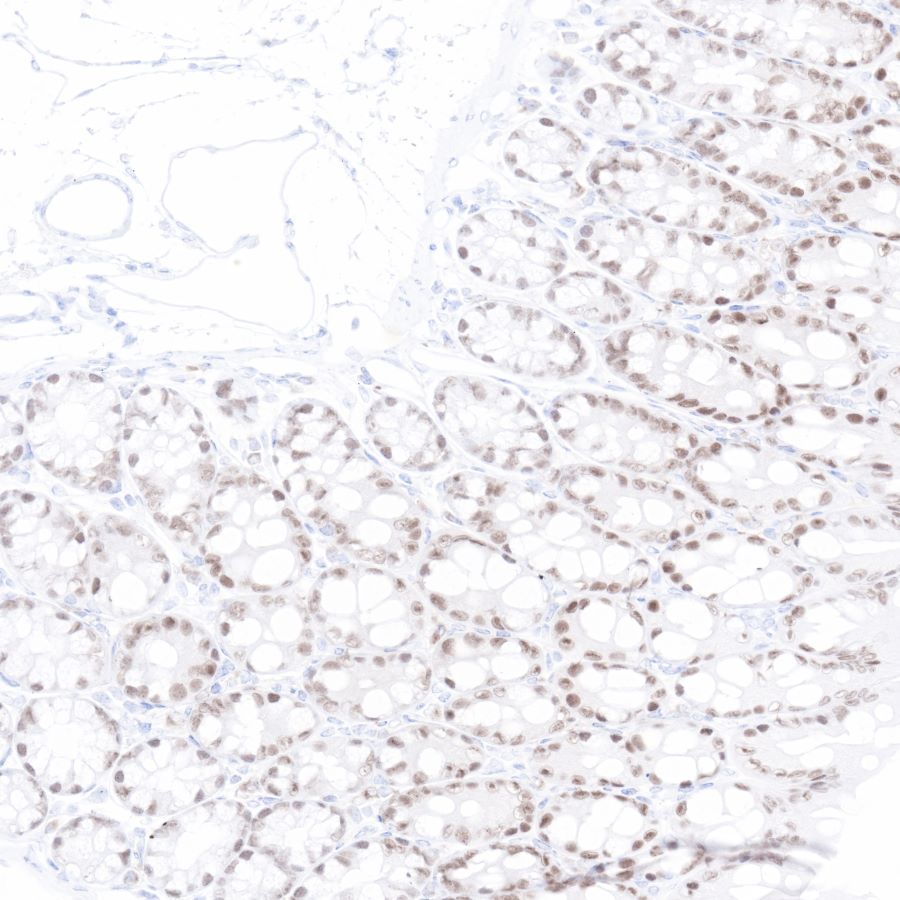
IHC shows positive staining in paraffin-embedded mouse colon.
Anti-CDX2 antibody was used at 1/500 dilution, followed by a Goat Anti-Rabbit IgG H&L (HRP) ready to use. Counterstained with hematoxylin.
Heat mediated antigen retrieval with Tris/EDTA buffer pH9.0 was performed before commencing with IHC staining protocol.
IHC shows positive staining in paraffin-embedded rat colon.
Anti-CDX2 antibody was used at 1/500 dilution, followed by a Goat Anti-Rabbit IgG H&L (HRP) ready to use. Counterstained with hematoxylin.
Heat mediated antigen retrieval with Tris/EDTA buffer pH9.0 was performed before commencing with IHC staining protocol.
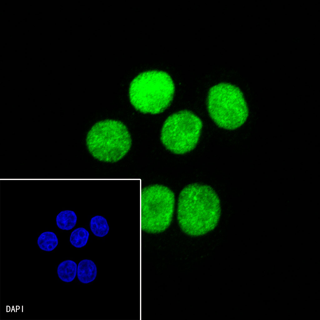
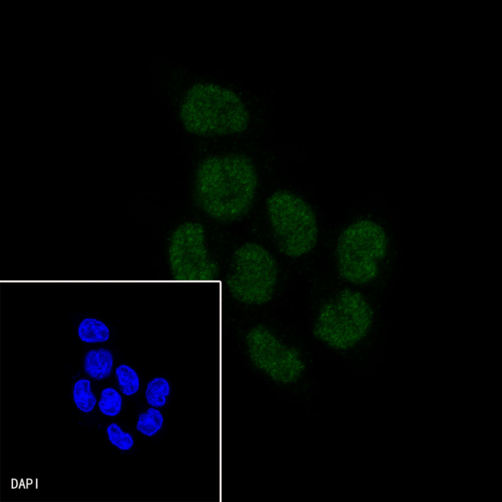
ICC shows positive nuclear staining in SW480 cells. Anti-CDX2 antibody was used at 1/200 dilution and incubated overnight at 4°C. Goat polyclonal Antibody to Rabbit IgG - H&L (Alexa Fluor® 488) was used as secondary antibody at 1/1000 dilution.The cells were fixed with 4% PFA and permeabilized with 0.1% PBS-Triton X-100. Nuclei were countersained with DAPI.

ICC shows positive nuclear staining in HCT-116 cells (Low expression). Anti-CDX2 antibody was used at 1/200 dilution and incubated overnight at 4°C. Goat polyclonal Antibody to Rabbit IgG - H&L (Alexa Fluor® 488) was used as secondary antibody at 1/1000 dilution.The cells were fixed with 4% PFA and permeabilized with 0.1% PBS-Triton X-100. Nuclei were countersained with DAPI.
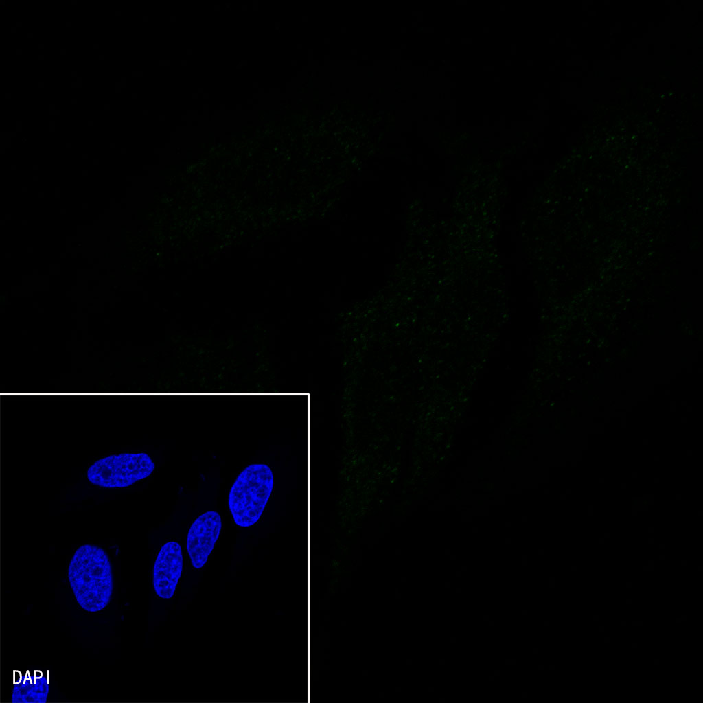
Negative control: ICC shows negative staining in HeLa cells. Anti-CDX2 antibody was used at 1/200 dilution and incubated overnight at 4°C. Goat polyclonal Antibody to Rabbit IgG - H&L (Alexa Fluor® 488) was used as secondary antibody at 1/1000 dilution.The cells were fixed with 4% PFA and permeabilized with 0.1% PBS-Triton X-100. Nuclei were countersained with DAPI.
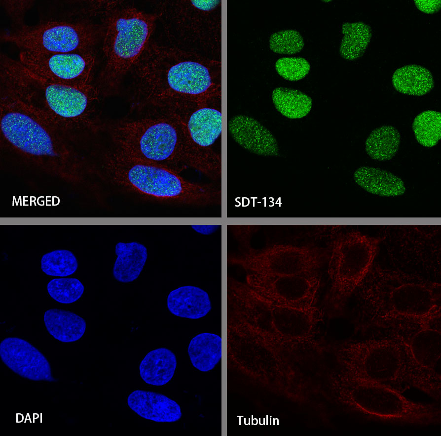
ICC shows positive staining in Caco-2 cells. Anti-CDX2 antibody was used at 1/500 dilution (Green) and incubated overnight at 4°C. Goat polyclonal Antibody to Rabbit IgG - H&L (Alexa Fluor® 488) was used as secondary antibody at 1/1000 dilution. The cells were fixed with 4%PFA and permeabilized with 0.1% PBS-Triton X-100. Nuclei were counterstained with DAPI (Blue). Counterstain with tubulin (red).
 您现在的位置: