
PBS, 40% Glycerol, 0.05%BSA, 0.03% Proclin 300
12 months from date of receipt / reconstitution, -20 °C as supplied
| 应用 | 稀释度 |
|---|---|
| IP | 1:25 |
| WB | 1:1000 |
| IHC-P | 1:500 |
The close connection consists of seal protein and closed protein, and can be connected to the cell skeleton. The Claudin family includes 23 integrated membrane proteins and their expression. Its expression is different from each tissue type, and can determine the intensity and characteristics of epithelial cell barrier. Claudin-1 is expressed in epithelial cells and nerve cysts.The change of Claudin protein expression mode is related to multiple cancer types. It mainly expresses the peripheral neuroma (partial type of negative), derivative skin fibrous sarcoma, low -level malignant fibrous mucus -like sarcoma, hardened fibroblastoma, fibroma, deity fibroma, and neurobetoma.
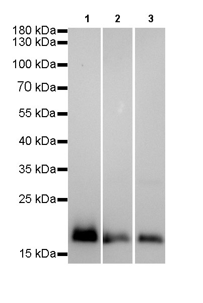
WB result of Claudin-1 Rabbit mAb
Primary antibody: Claudin-1 Rabbit mAb at 1/1000 dilution
Lane 1:A431 whole cell lysate 20 µg
Lane 2:A549 whole cell lysate 20 µg
Lane 3:HepG2 whole cell lysate 20 µg
low expression control: A549 whole cell lysate
Secondary antibody: Goat Anti-Rabbit IgG, (H+L), HRP conjugated at 1/10000 dilution
Predicted MW: 20 kDa
Observed MW: 20 kDa
Exposure time: Lane 1: 1.5s
Lane 2 and Lane 3: 60s

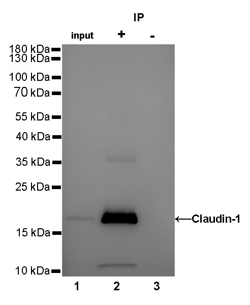
Claudin-1 Rabbit mAb at 1/25 dilution (2µg) immunoprecipitating Claudin-1 in 0.4mg A431 whole cell lysate. Western blot was performed on the immunoprecipitate using Claudin-1 Rabbit mAb at 1/1000 dilution. Secondary antibody (HRP) for IP was used at 1/400 dilution.
Lane 1: A431 whole cell lysate 10µg (input)
Lane 2: Claudin-1 Rabbit mAb IP in A431 whole cell lysate
Lane 3: Rabbit monoclonal IgG IP in A431 whole cell lysate
Predicted MW: 20 kDa
Observed MW: 20 kDa
Exposure time: 2.4s
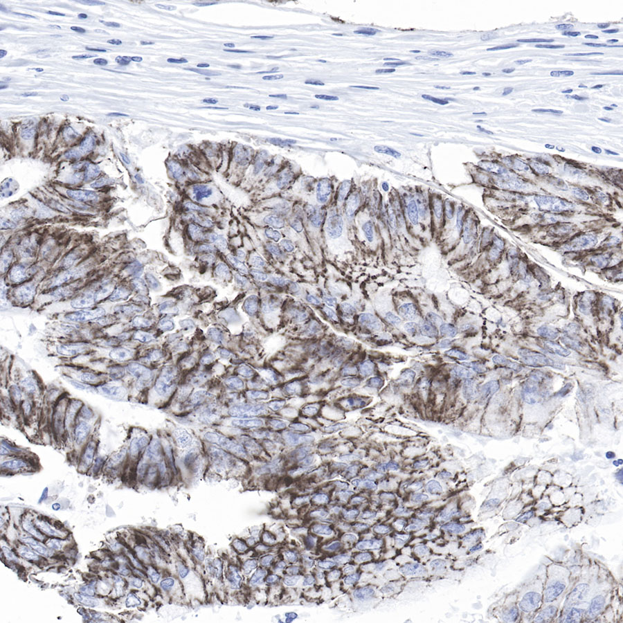
IHC shows positive staining in paraffin-embedded human colon cancer.
Anti-Claudin-1 antibody was used at 1/500 dilution, followed by a Goat Anti-Rabbit IgG H&L (HRP) ready to use. Counterstained with hematoxylin.
Heat mediated antigen retrieval with Tris/EDTA buffer pH9.0 was performed before commencing with IHC staining protocol.
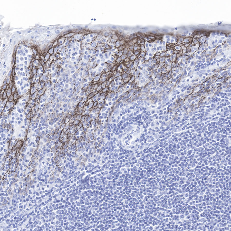
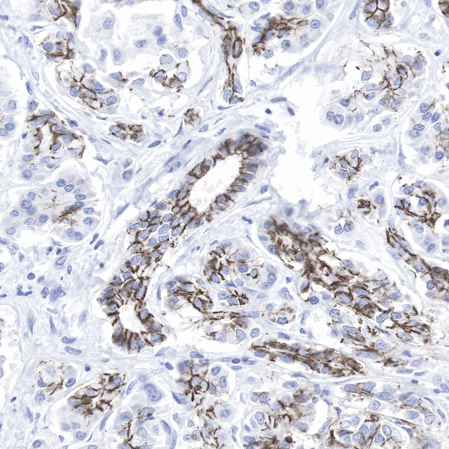
IHC shows positive staining in paraffin-embedded human tonsil.
Anti-Claudin-1 antibody was used at 1/500 dilution, followed by a Goat Anti-Rabbit IgG H&L (HRP) ready to use. Counterstained with hematoxylin.
Heat mediated antigen retrieval with Tris/EDTA buffer pH9.0 was performed before commencing with IHC staining protocol.
IHC shows positive staining in paraffin-embedded human skin.
Anti-Claudin-1 antibody was used at 1/500 dilution, followed by a Goat Anti-Rabbit IgG H&L (HRP) ready to use. Counterstained with hematoxylin.
Heat mediated antigen retrieval with Tris/EDTA buffer pH9.0 was performed before commencing with IHC staining protocol.
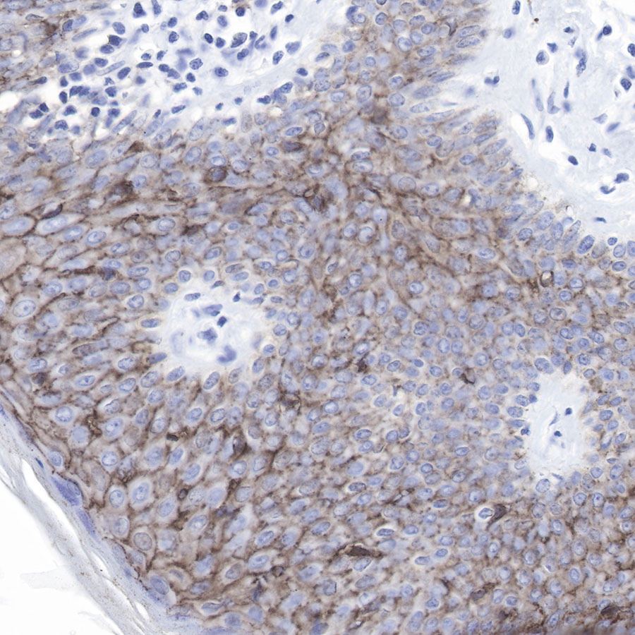
IHC shows positive staining in paraffin-embedded human pancreas cancer.
Anti-Claudin-1 antibody was used at 1/500 dilution, followed by a Goat Anti-Rabbit IgG H&L (HRP) ready to use. Counterstained with hematoxylin.
Heat mediated antigen retrieval with Tris/EDTA buffer pH9.0 was performed before commencing with IHC staining protocol.
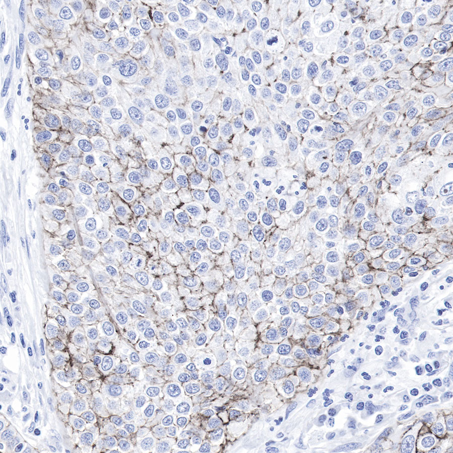
IHC shows positive staining in paraffin-embedded human lung squamous cancer.
Anti-Claudin-1 antibody was used at 1/500 dilution, followed by a Goat Anti-Rabbit IgG H&L (HRP) ready to use. Counterstained with hematoxylin.
Heat mediated antigen retrieval with Tris/EDTA buffer pH9.0 was performed before commencing with IHC staining protocol.
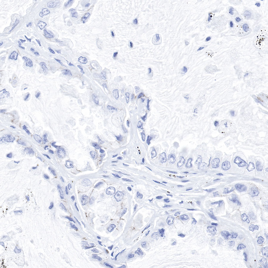
Negative control.IHC shows negative staining in paraffin-embedded human lung adenocarcinoma cancer.
Anti-Claudin-1 antibody was used at 1/500 dilution, followed by a Goat Anti-Rabbit IgG H&L (HRP) ready to use. Counterstained with hematoxylin.
Heat mediated antigen retrieval with Tris/EDTA buffer pH9.0 was performed before commencing with IHC staining protocol.
 您现在的位置: