
PBS, 40% Glycerol, 0.05%BSA, 0.03% Proclin 300
12 months from date of receipt / reconstitution, -20 °C as supplied
| 应用 | 稀释度 |
|---|---|
| WB | 1:1000-1:5000 |
| IHC-P | 1:2000 |
Tumor protein p63, typically referred to as p63, also known as transformation-related protein 63 is a protein that in humans is encoded by the TP63 (also known as the p63) gene. Transcription factor p63 is a key regulator of epidermal keratinocyte proliferation and differentiation. Tumor protein p63 is a member of the p53 family of transcription factors. p63 regulates the activity of a multitude of genes involved in growth and development of the ectoderm and derived structures and tissues, such as basal layer keratins and cell cycle control genes. Accordingly, p63 expression is found in basal cell layers of various organs, squamous epithelial cells of many organs and urothelium. p63 (TAp63) is closely related to p40 (ΔNp63) as both proteins represent isoforms of the p63 gene with distinct molecular functions. While “full length” p63 (TAp63) activates p53 target genes such as p21 or BAX, the shorter transcript p40 (ΔNp63) inhibits activation of p53 and “full length” p63. P63 is also helpful in distinguishing poorly differentiated squamous cell carcinoma from small cell carcinoma or adenocarcinoma. P63 should be strongly stained in poorly differentiated squamous cell, but negative in small cell or adenocarcinoma.
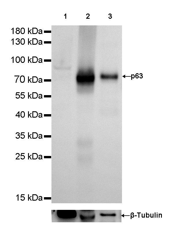
WB result of p63 Rabbit mAb
Primary antibody: p63 Rabbit mAb at 1/1000 dilution
Lane 1: MCF-7 whole cell lysate 20ug
Lane 2: Hacat whole cell lysate 20ug
Lane 3: A431 whole cell lysate 20ug
Negative control: MCF-7 whole cell lysate
Secondary antibody: Goat Anti-Rabbit IgG (H+L), HRP conjugated at 1/10000 dilution
Predicted MW: 76 kDa
Observed MW: 76 kDa
Exposure time: 1 seconds
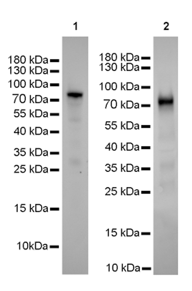
WB result of p63 Rabbit mAb
Primary antibody: p63 Rabbit mAb at 1/1000 dilution
Lane 1: mouse skin whole lysate 20ug
Lane 2: mouse bladder whole lysate 20ug
Secondary antibody: Goat Anti-Rabbit IgG (H+L), HRP conjugated at 1/10000 dilution
Predicted MW: 76 kDa
Observed MW: 76 kDa
Exposure time: lane 1: 60 seconds; lane 2: 120 seconds
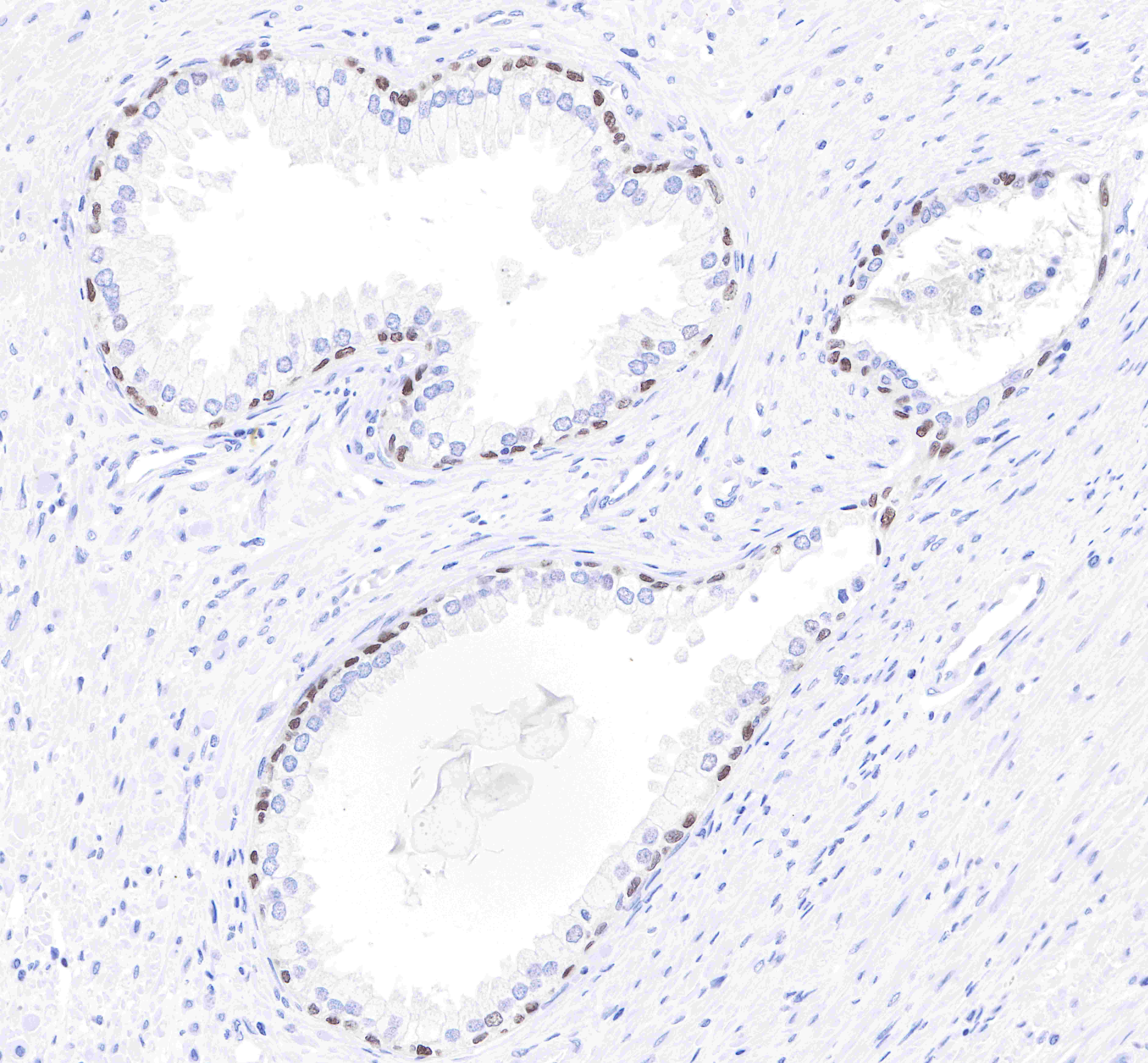
IHC shows positive staining in paraffin-embedded human prostate.
Anti-p63 antibody was used at 1/2000 dilution, followed by a Goat Anti-Rabbit IgG H&L (HRP) ready to use.
Counterstained with hematoxylin.
Heat mediated antigen retrieval with Tris/EDTA buffer pH9.0 was performed before commencing with IHC staining protocol.

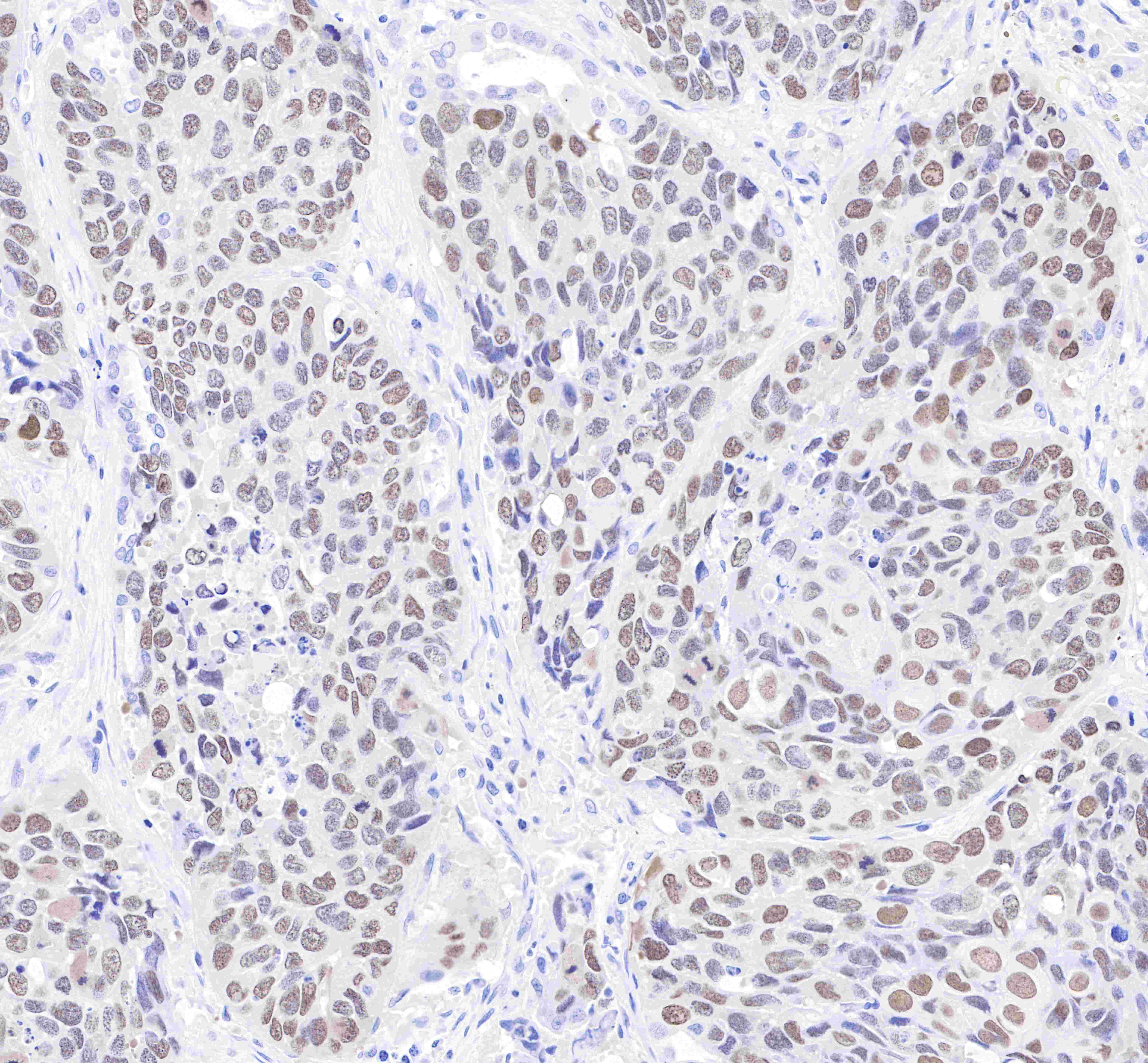
IHC shows positive staining in paraffin-embedded human lung squamous cell carcinoma.
Anti-p63 antibody was used at 1/2000 dilution, followed by a Goat Anti-Rabbit IgG H&L (HRP) ready to use.
Counterstained with hematoxylin.
Heat mediated antigen retrieval with Tris/EDTA buffer pH9.0 was performed before commencing with IHC staining protocol.
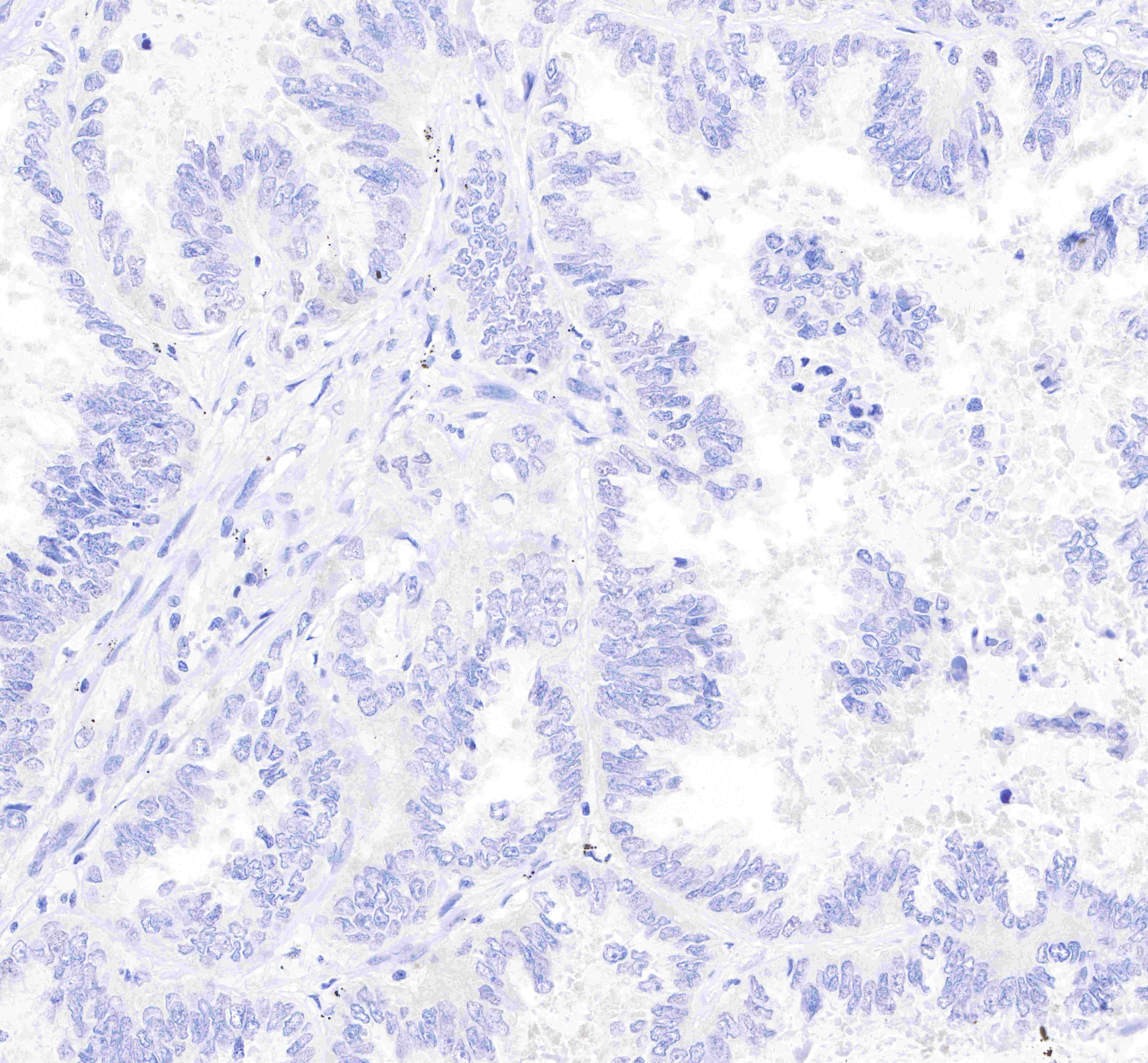
Negative control: IHC shows negative staining in paraffin-embedded human lung adenocarcinoma.
Anti-p63 antibody was used at 1/2000 dilution, followed by a Goat Anti-Rabbit IgG H&L (HRP) ready to use.
Counterstained with hematoxylin.
Heat mediated antigen retrieval with Tris/EDTA buffer pH9.0 was performed before commencing with IHC staining protocol.
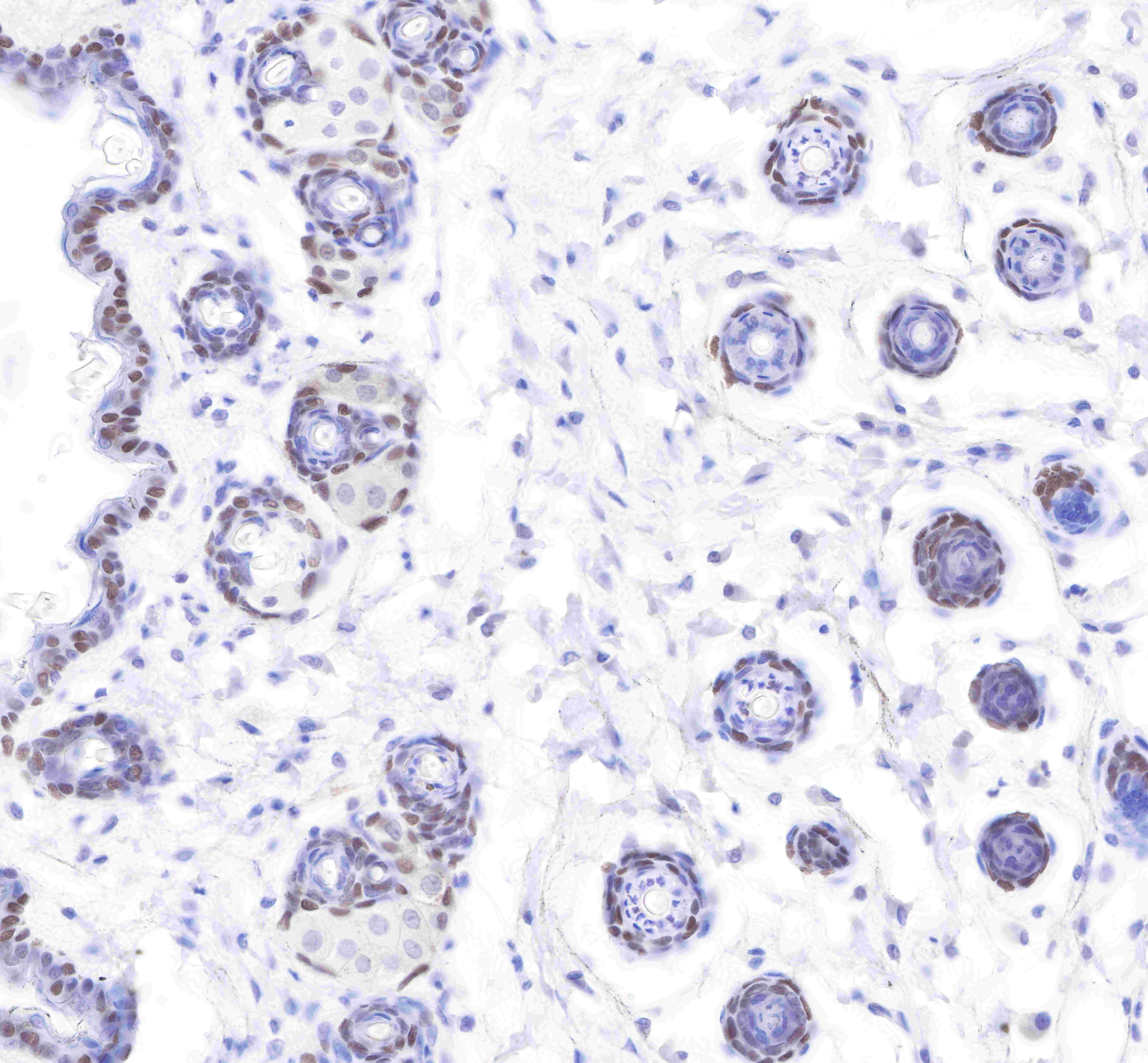
IHC shows positive staining in paraffin-embedded mouse skin.
Anti-p63 antibody was used at 1/2000 dilution, followed by a Goat Anti-Rabbit IgG H&L (HRP) ready to use.
Counterstained with hematoxylin.
Heat mediated antigen retrieval with Tris/EDTA buffer pH9.0 was performed before commencing with IHC staining protocol.
 您现在的位置: