
| 应用 | 稀释度 |
|---|---|
| IHC-P | 1:500 |
| ICC | 1:500 |
| IP | 1:25 |
| WB | 1:1000 |
| FC | 1:500 |
SATB1, the global chromatin organizer and transcription factor, has emerged as a key factor integrating higher-order chromatin architecture with gene regulation. Recent studies have unraveled the role of SATB1 in organization of chromatin 'loopscape' and its dynamic nature in response to physiological stimuli. At genome-wide level, SATB1 seems to play a role in organization of the transcriptionally poised chromatin. SATB1 organizes the MHC class-I locus into distinct chromatin loops by tethering MARs to nuclear matrix at fixed distances. Silencing of SATB1 mimics the effects of IFN-γ treatment on chromatin loop architecture of the MHC class I locus and altered expression of genes within the locus. SATB1 has also been shown to induce breast cancer tumor growth and metastasis through the altered expression of large numbers of genes.

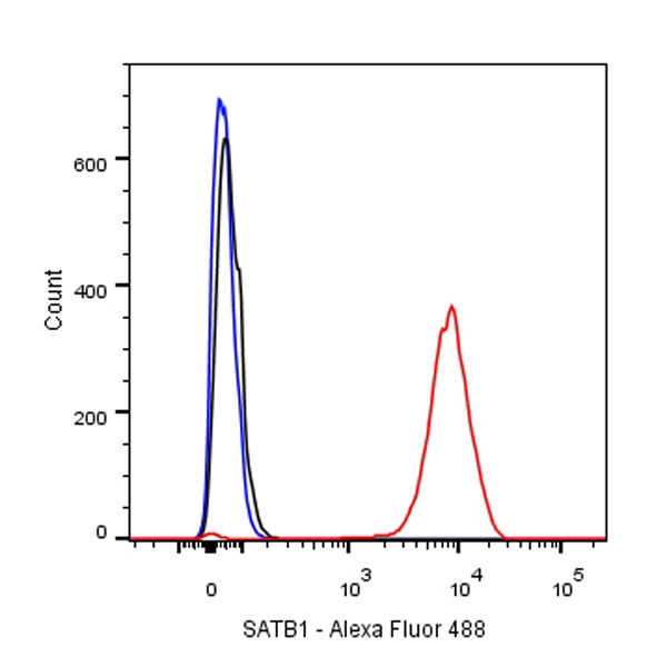
Flow cytometric analysis of Jurkat cells labelling SATB1 antibody at 1/500 dilution (0.1ug)/ (red) compared with a Rabbit monoclonal IgG (Black) isotype control and an unlabelled control (cells without incubation with primary antibody and secondary antibody) (Blue).
Goat Anti-Rabbit IgG Alexa Fluor® 488 at 1/1000 dilution was used as the secondary antibody.
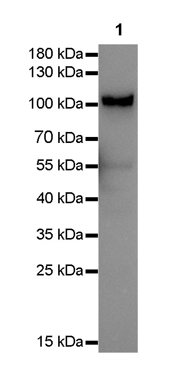
WB result of SATB1 Rabbit mAb
Primary antibody: SATB1 Rabbit mAb at 1/1000 dilution
Lane 1: Jurkat whole cell lysate 20 µg
Secondary antibody: Goat Anti-Rabbit IgG, (H+L), HRP conjugated at 1/10000 dilution
Predicted MW: 100 kDa
Observed MW: 100 kDa
Exposure time: 1s
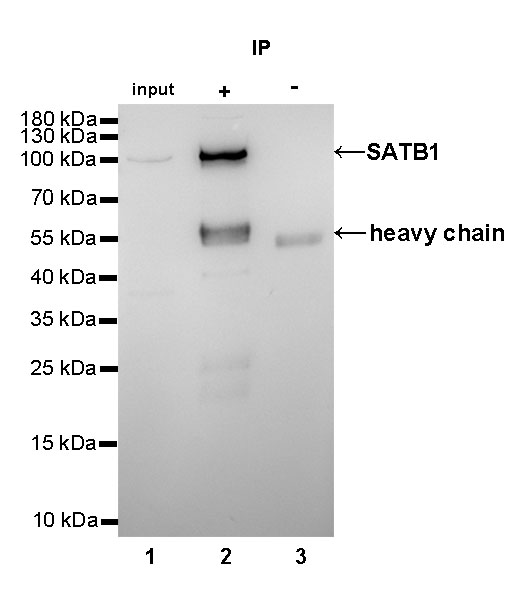
SATB1 Rabbit mAb at 1/25 dilution (2µg) immunoprecipitating SATB1 in 0.4mg Jurkat whole cell lysate.
Western blot was performed on the immunoprecipitate using SATB1 Rabbit mAb at 1/1000 dilution.
Secondary antibody (HRP) for IP was used at 1/400 dilution.
Lane 1: Jurkat whole cell lysate 10µg (input)
Lane 2 (+): SATB1 Rabbit mAb IP in Jurkat whole cell lysate
Lane 3 (-): Rabbit monoclonal IgG IP in Jurkat whole cell lysate
Predicted MW: 100 kDa
Observed MW: 100 kDa
Exposure time: 10s
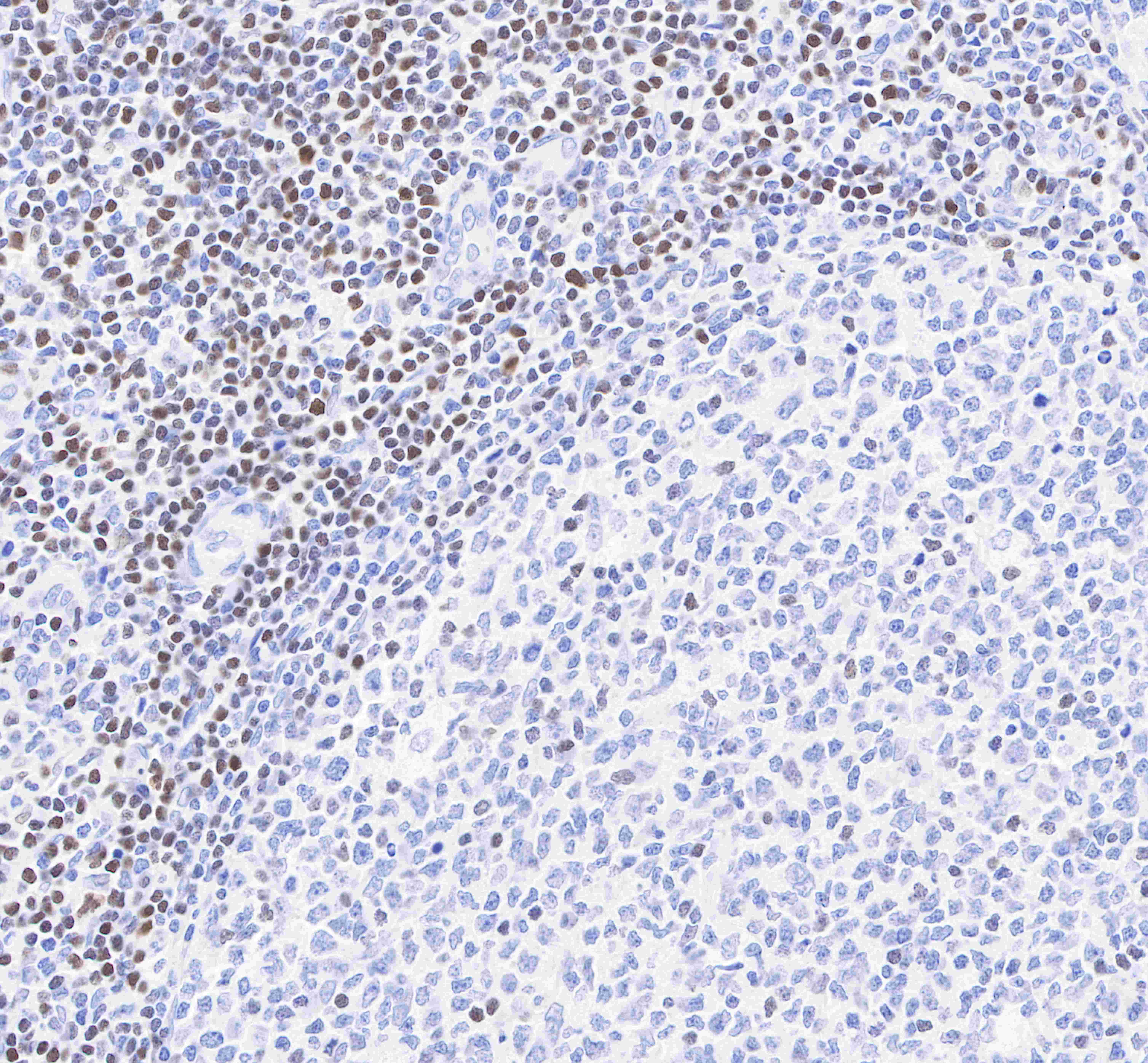
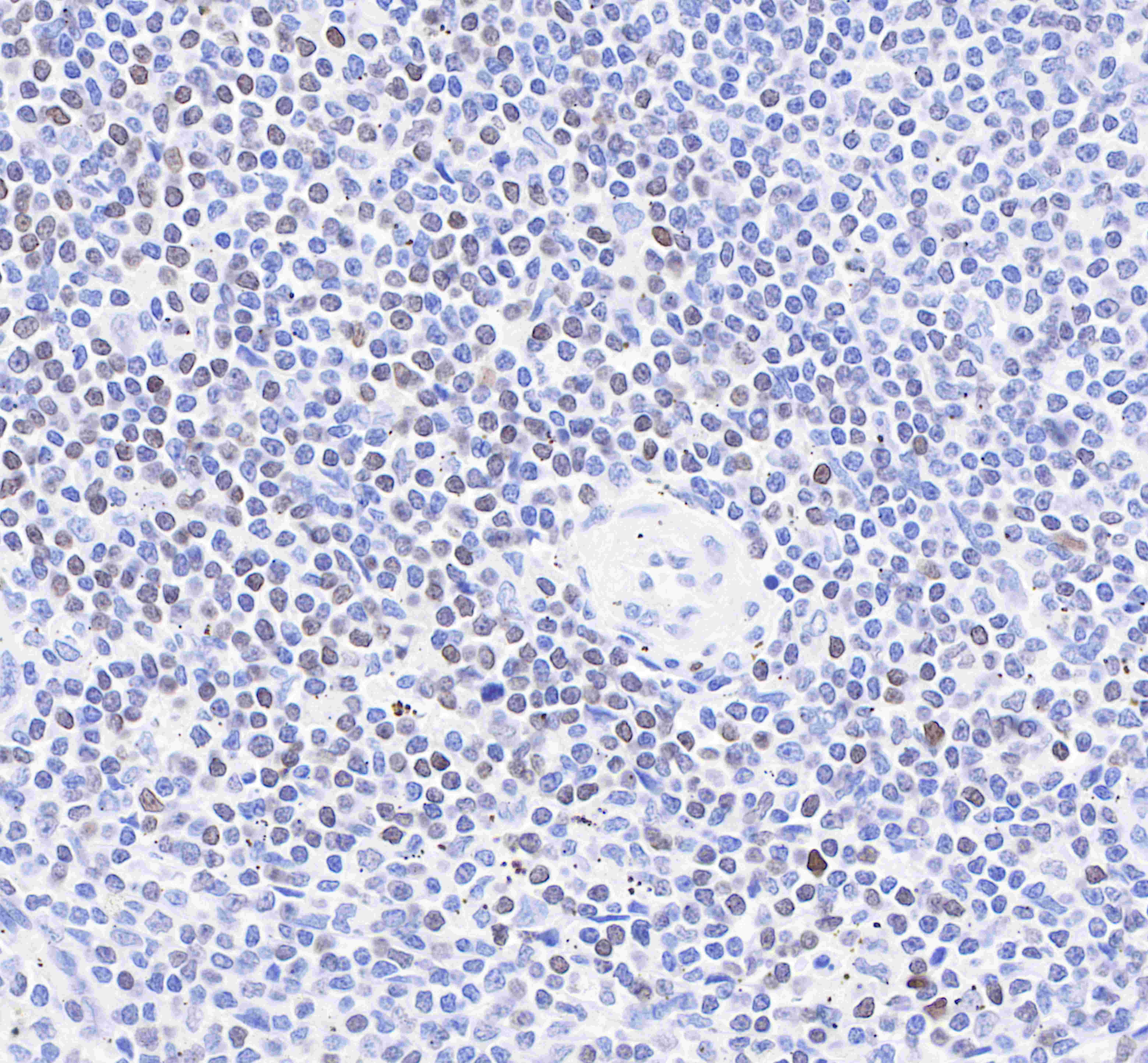
IHC shows positive staining in paraffin-embedded human tonsil.
Anti-SATB1 antibody was used at 1/500 dilution, followed by a Goat Anti-Rabbit IgG H&L (HRP) ready to use. Counterstained with hematoxylin.
Heat mediated antigen retrieval with Tris/EDTA buffer pH9.0 was performed before commencing with IHC staining protocol.
IHC shows positive staining in paraffin-embedded human spleen.
Anti-SATB1 antibody was used at 1/500 dilution, followed by a Goat Anti-Rabbit IgG H&L (HRP) ready to use. Counterstained with hematoxylin.
Heat mediated antigen retrieval with Tris/EDTA buffer pH9.0 was performed before commencing with IHC staining protocol.
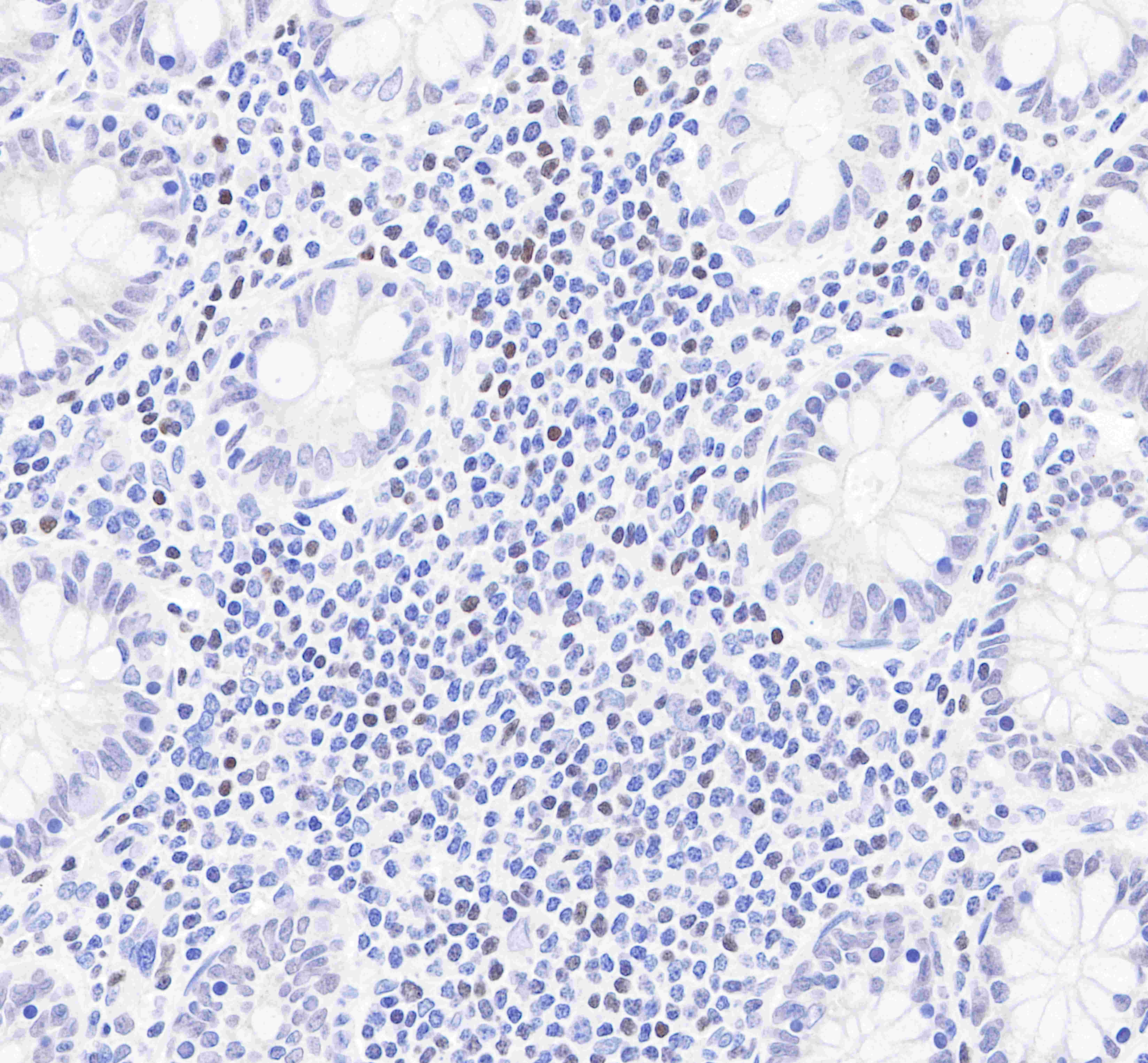
IHC shows positive staining in paraffin-embedded human colon.
Anti-SATB1 antibody was used at 1/500 dilution, followed by a Goat Anti-Rabbit IgG H&L (HRP) ready to use. Counterstained with hematoxylin.
Heat mediated antigen retrieval with Tris/EDTA buffer pH9.0 was performed before commencing with IHC staining protocol.
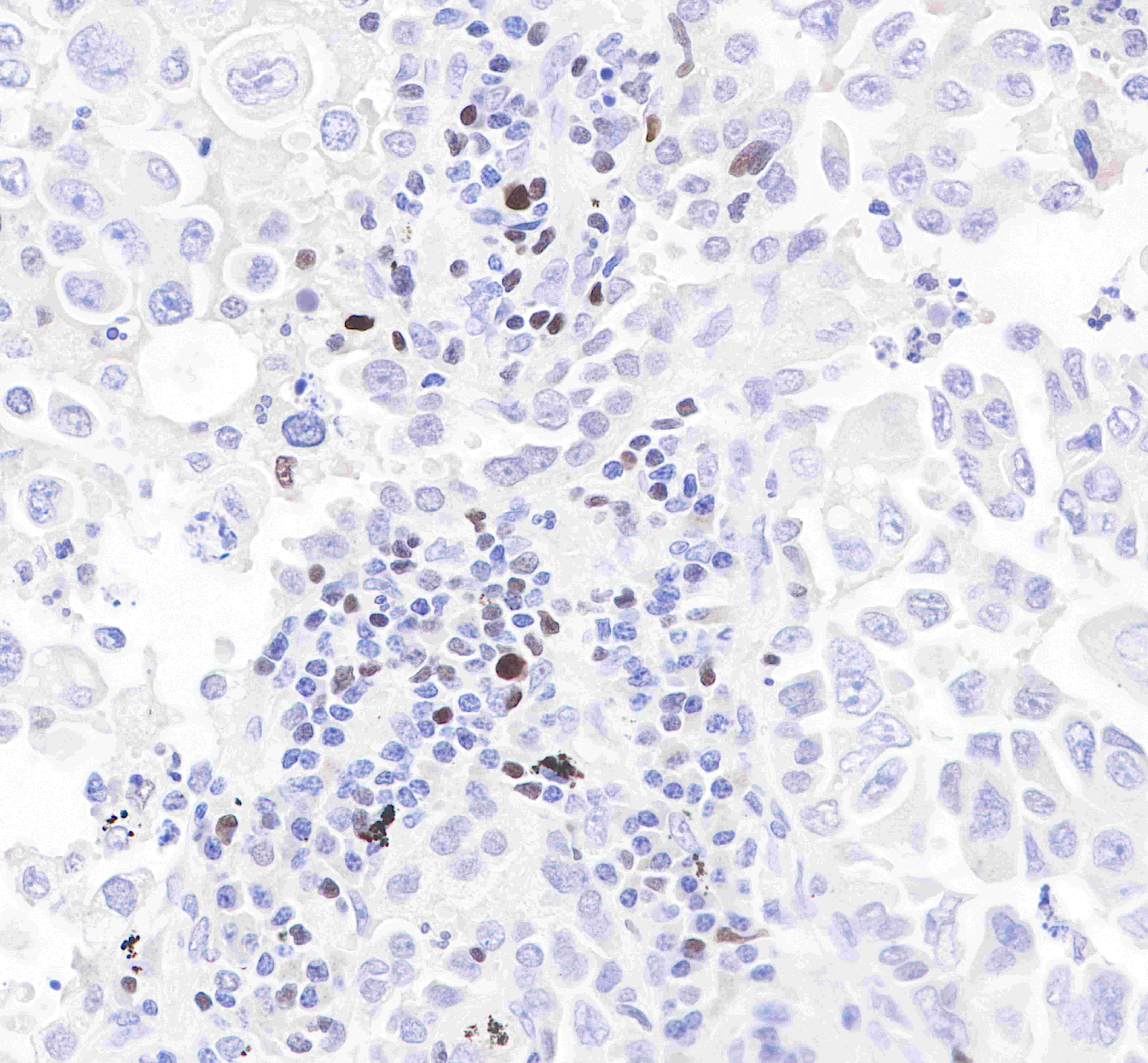
IHC shows positive staining in paraffin-embedded human lung adenocarcinoma.
Anti-SATB1 antibody was used at 1/500 dilution, followed by a Goat Anti-Rabbit IgG H&L (HRP) ready to use. Counterstained with hematoxylin.
Heat mediated antigen retrieval with Tris/EDTA buffer pH9.0 was performed before commencing with IHC staining protocol.
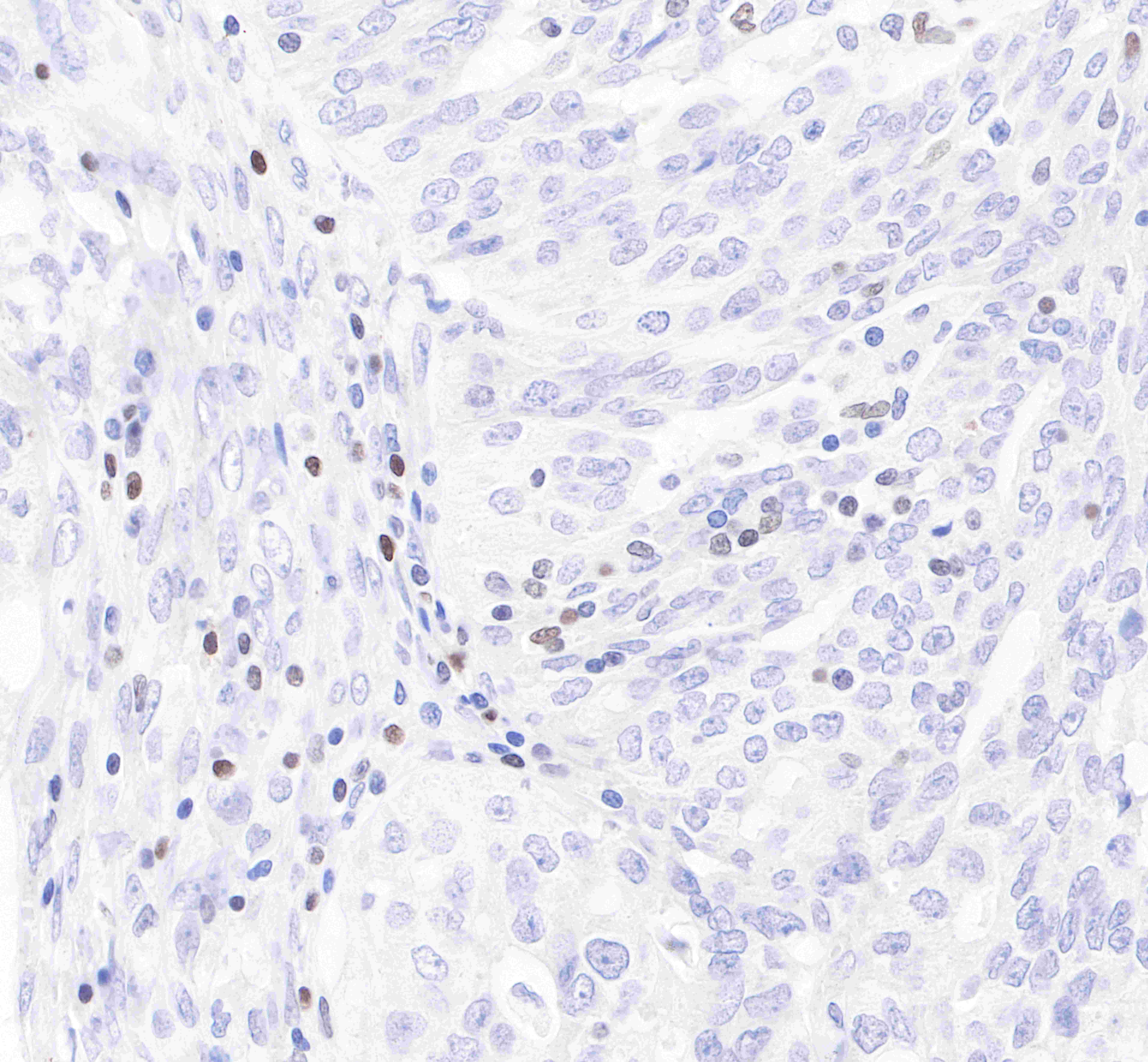
IHC shows positive staining in paraffin-embedded human lung ovarian cancer.
Anti-SATB1 antibody was used at 1/500 dilution, followed by a Goat Anti-Rabbit IgG H&L (HRP) ready to use. Counterstained with hematoxylin.
Heat mediated antigen retrieval with Tris/EDTA buffer pH9.0 was performed before commencing with IHC staining protocol.
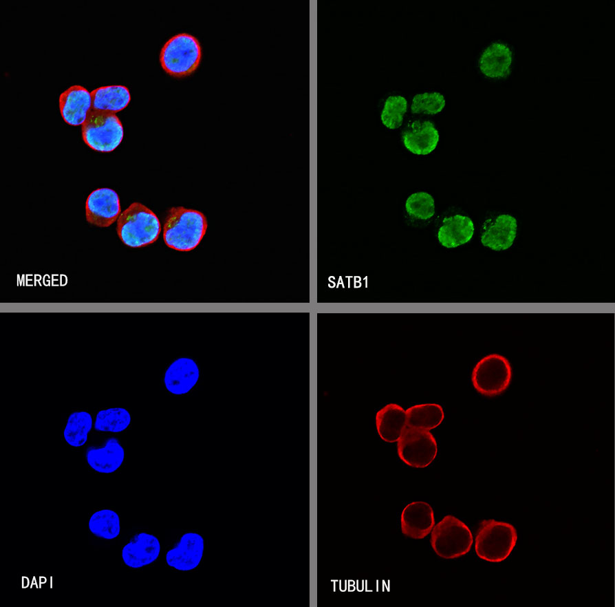
ICC shows positive staining in Jurkat cells. Anti-SATB1 antibody was used at 1/500 dilution (Green) and incubated overnight at 4°C. Goat polyclonal Antibody to Rabbit IgG - H&L (Alexa Fluor® 488) was used as secondary antibody at 1/1000 dilution. The cells were fixed with 4% PFA and permeabilized with 0.1% PBS-Triton X-100. Nuclei were counterstained with DAPI (Blue).Counterstain with tubulin (Red).
 您现在的位置: