
| 应用 | 稀释度 |
|---|---|
| ICFCM | 1:500 |
| ICC | 1:500 |
| IHC-P | 1: 2000 |
| IP | 1:25 |
| WB | 1:2000 |
DNA replication licensing factor MCM3 is a protein that in humans is encoded by the MCM3 gene. The protein encoded by this gene is one of the highly conserved mini-chromosome maintenance proteins (MCM) that are involved in the initiation of eukaryotic genome replication. The hexameric protein complex formed by MCM proteins is a key component of the pre-replication complex (pre-RC) and may be involved in the formation of replication forks and in the recruitment of other DNA replication related proteins. This protein is a subunit of the protein complex that consists of MCM2-7. It has been shown to interact directly with MCM5/CDC46. This protein also interacts with, and thus is acetylated by MCM3AP, a chromatin-associated acetyltransferase. The acetylation of this protein inhibits the initiation of DNA replication and cell cycle progression.

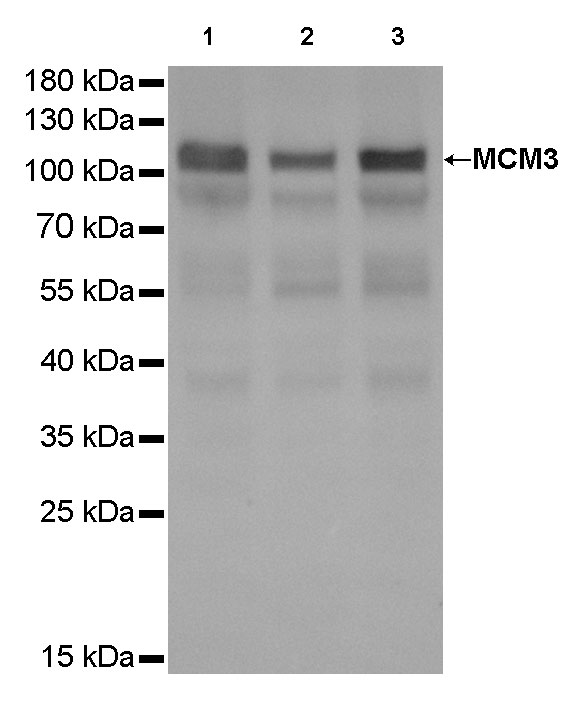
WB result of MCM3 Rabbit mAb
Primary antibody : MCM3 Rabbit mAb at 1/2000 dilution
Lane 1: Hela whole cell lysate 20 µg
Lane 2: HepG2 whole cell lysate 20 µg
Lane 3: K562 whole cell lysate 20 µg
Secondary antibody: Goat Anti-Rabbit IgG, (H+L), HRP conjugated at 1/10000 dilution
Predicted MW: 91 kDa
Observed MW: 110 kDa
Exposure time: 1.3 s
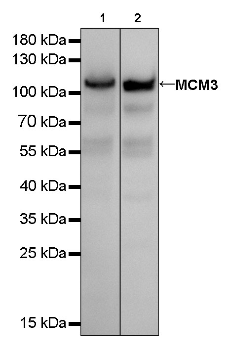
WB result of MCM3 Rabbit mAb
Primary antibody : MCM3 Rabbit mAb at 1/2000 dilution
Lane 1: mouse spleen lysate 20 µg
Lane 2: NIH/3T3 whole cell lysate 20 µg
Secondary antibody: Goat Anti-Rabbit IgG, (H+L), HRP conjugated at 1/10000 dilution
Predicted MW: 91 kDa
Observed MW: 110 kDa
Exposure time: 4 s
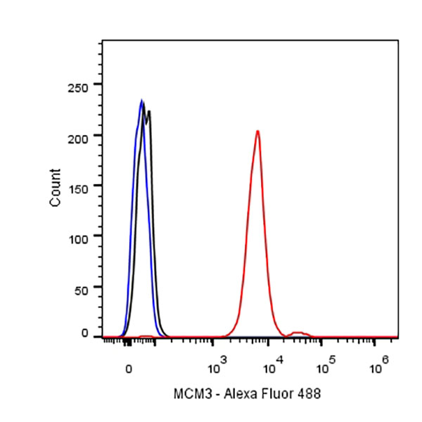
Flow cytometric analysis of A431 cells labelling MCM3 antibody at 1/500 (0.1ug) dilution/ (red) compared with a Rabbit monoclonal IgG (Black) isotype control and an unlabelled control (cells without incubation with primary antibody and secondary antibody) (Blue). Goat Anti-Rabbit IgG Alexa Fluor® 488 was used as the secondary antibody.
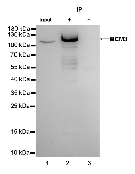
MCM3 Rabbit mAb at 1/25 dilution (2µg) immunoprecipitating MCM3 in 0.4mg HeLa whole cell lysate. Western blot was performed on the immunoprecipitate using MCM3 Rabbit mAb at 1/1000 dilution. Secondary antibody (HRP) for IP was used at 1/400 dilution.
Lane 1: HeLa whole cell lysate 10µg (input)
Lane 2: MCM3 Rabbit mAb IP in HeLa whole cell lysate
Lane 3: Rabbit monoclonal IgG IP in HeLa whole cell lysate
Predicted MW: 91 kDa
Observed MW: 110 kDa
Exposure time: 15s
IHC shows positive staining in paraffin-embedded human stomach.
Anti-MCM3 antibody was used at 1/2000 dilution, followed by a Goat Anti-Rabbit IgG H&L (HRP) ready to use.
Counterstained with hematoxylin.
Heat mediated antigen retrieval with Tris/EDTA buffer pH9.0 was performed before commencing with IHC staining protocol.
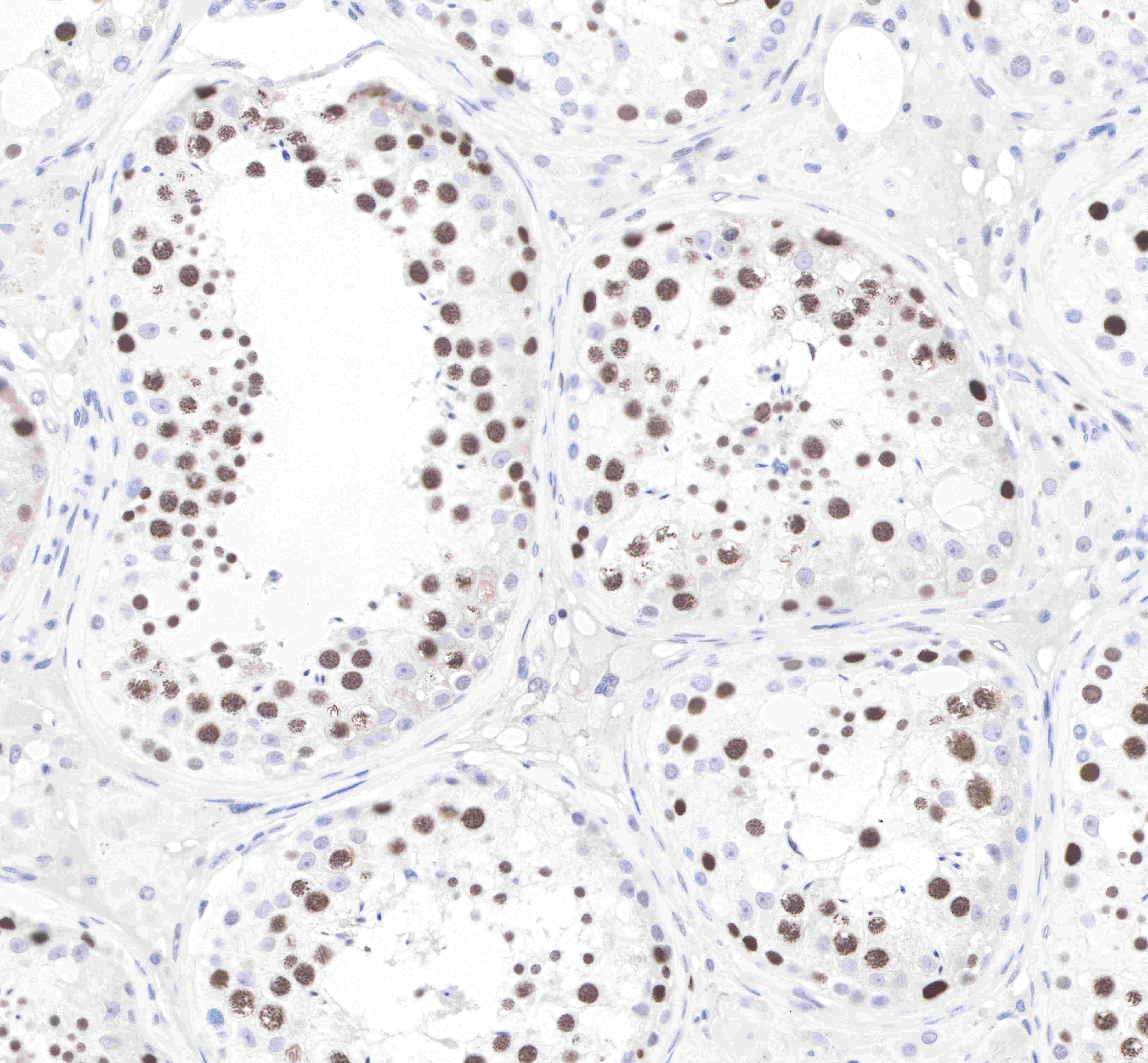
IHC shows positive staining in paraffin-embedded human testis. Anti-MCM3 antibody was used at 1/2000 dilution, followed by a Goat Anti-Rabbit IgG H&L (HRP) ready to use.
Counterstained with hematoxylin.
Heat mediated antigen retrieval with Tris/EDTA buffer pH9.0 was performed before commencing with IHC staining protocol.
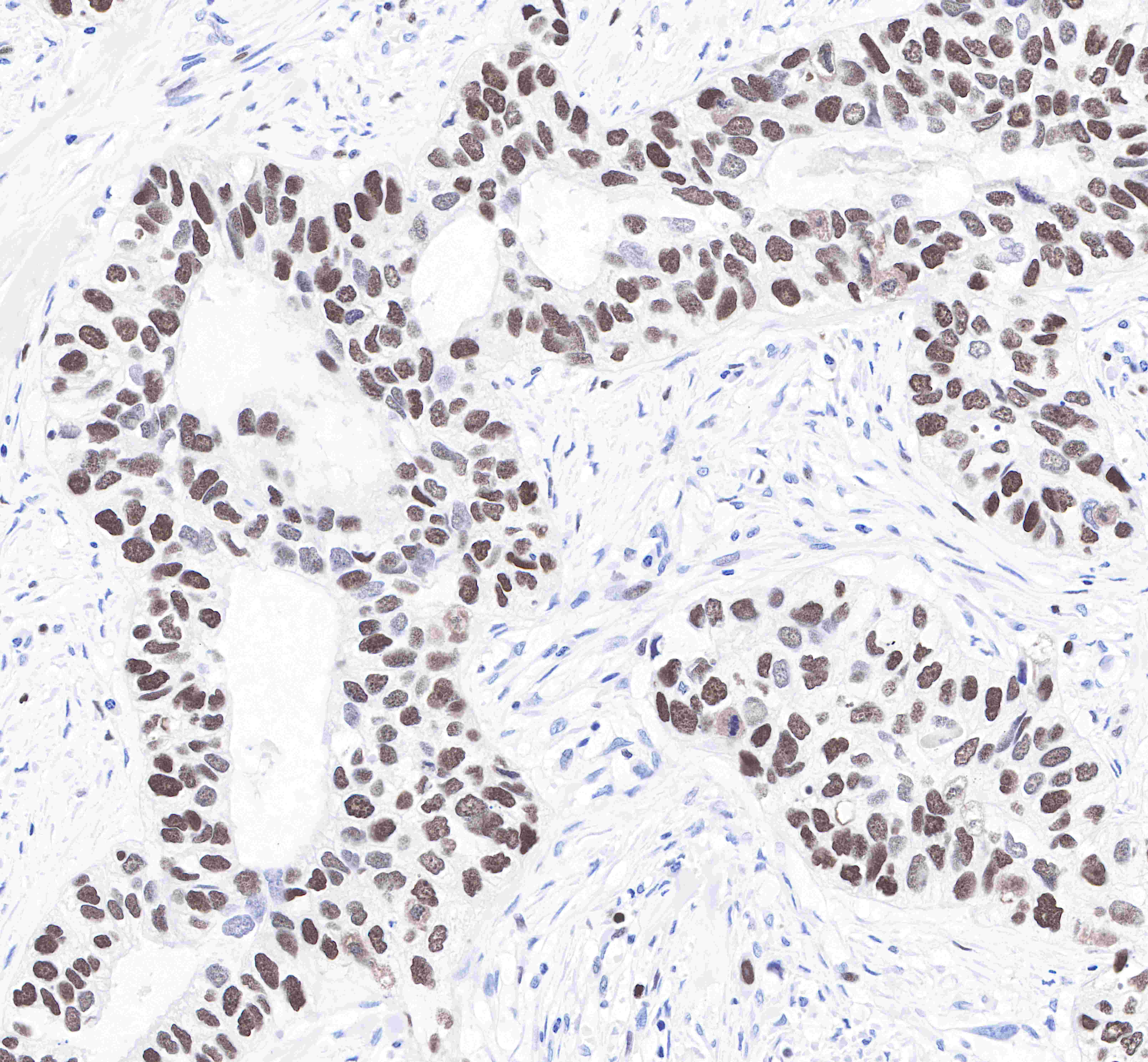
IHC shows positive staining in paraffin-embedded human breast cancer.
Anti-MCM3 antibody was used at 1/2000 dilution, followed by a Goat Anti-Rabbit IgG H&L (HRP) ready to use.
Counterstained with hematoxylin.
Heat mediated antigen retrieval with Tris/EDTA buffer pH9.0 was performed before commencing with IHC staining protocol.
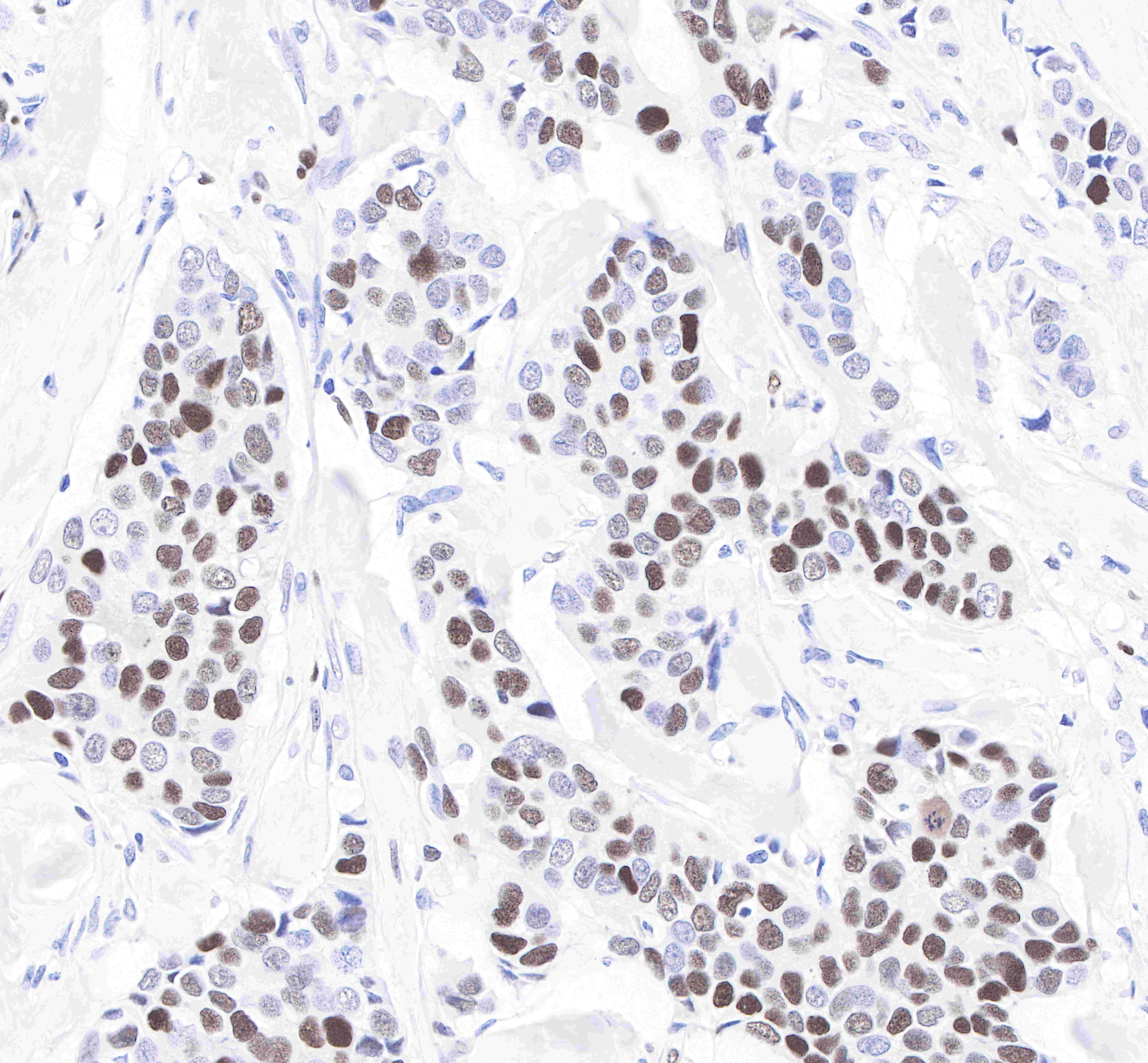
IHC shows positive staining in paraffin-embedded human lung squamous cell cancer.
Anti-MCM3 antibody was used at 1/2000 dilution, followed by a Goat Anti-Rabbit IgG H&L (HRP) ready to use.
Counterstained with hematoxylin.
Heat mediated antigen retrieval with Tris/EDTA buffer pH9.0 was performed before commencing with IHC staining protocol.
IHC shows positive staining in paraffin-embedded mouse testis. Anti-MCM3 antibody was used at 1/2000 dilution, followed by a Goat Anti-Rabbit IgG H&L (HRP) ready to use.
Counterstained with hematoxylin.
Heat mediated antigen retrieval with Tris/EDTA buffer pH9.0 was performed before commencing with IHC staining protocol.
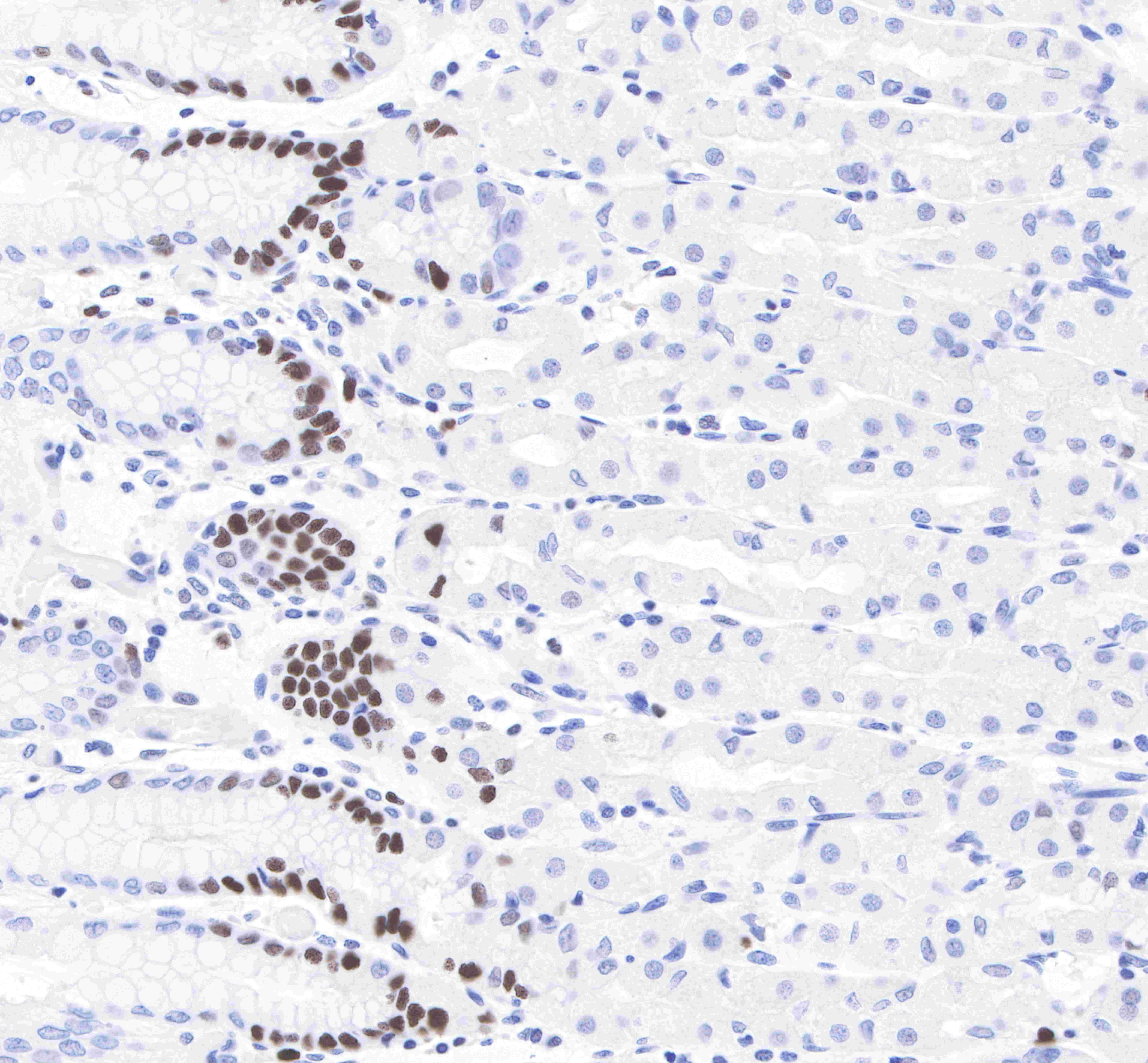
IHC shows positive staining in paraffin-embedded rat spleen.
Anti-MCM3 antibody was used at 1/2000 dilution, followed by a Goat Anti-Rabbit IgG H&L (HRP) ready to use.
Counterstained with hematoxylin.
Heat mediated antigen retrieval with Tris/EDTA buffer pH9.0 was performed before commencing with IHC staining protocol.
ICC shows nuclear staining in HeLa cells.
Anti-MCM3 antibody was used at 1/500 dilution and incubated overnight at 4°C. Goat polyclonal Antibody to Rabbit IgG - H&L (Alexa Fluor® 488) was used as secondary antibody at 1/1000 dilution (shown in green).
The cells were fixed with 100% methanol and permeabilized with 0.1% PBS-Triton X-100. Nuclei were counterstained with DAPI.
 您现在的位置: