
12 months from date of receipt / reconstitution, -20 °C as supplied
| 应用 | 稀释度 |
|---|---|
| IHC-P | 1:2000 |
| WB | 1:1000 |
| ICFCM | 1:5000 |
| ICC | 1:500 |
Annexin A1 belongs to the annexin family of Ca2+-dependent phospholipid-binding proteins that have a molecular weight of approximately 35,000 to 40,000 Dalton and are preferentially located on the cytosolic face of the plasma membrane. Annexin A1 protein has an apparent relative molecular mass of 40 kDa with phospholipase A2 inhibitory activity. In resting conditions, human and mouse immune cells such as neutrophils, monocytes, and macrophages contain high levels of annexin A1 in their cytoplasm. Following cell activation (for example, by neutrophil adhesion to endothelial-cell monolayers), annexin A1 is promptly mobilized to the cell surface and secreted. Annexin A1 promotes neutrophil detachment and apoptosis, and phagocytosis of apoptotic neutrophils by macrophages. On the other hand, it reduces the tendency of neutrophils to penetrate the endothelium of blood vessels. Higher expression of annexin A1 during pathological conditions could increase the strength of TCR signalling through the mitogen-activated protein kinase signalling pathway, thereby causing a state of hyperactivation of T cells.

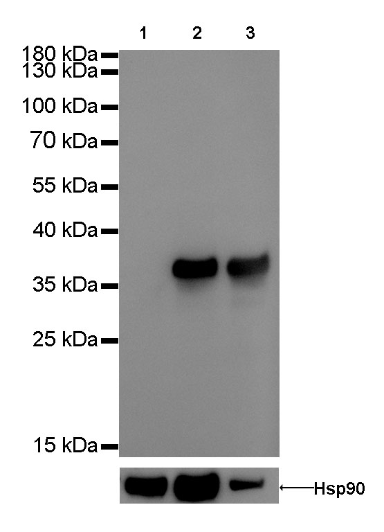
WB result of Annexin A1 Rabbit mAb
Primary antibody : Annexin A1 Rabbit mAb at 1/1000 dilution
Lane 1: 293T whole cell lysate 20 µg
Lane 2: K562 whole cell lysate 20 µg
Lane 3: A549 whole cell lysate 20 µg
Negative control: 293T whole cell lysate
Secondary antibody: Goat Anti-Rabbit IgG, (H+L), HRP conjugated at 1/10000 dilution
Predicted MW: 38 kDa
Observed MW: 38 kDa
Exposure time: 1s
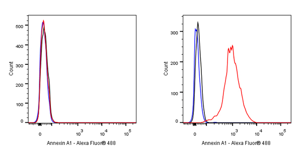
Flow cytometric analysis of 4% PFA fixed 90% methanol permeabilized 293T (Human embryonic kidney epithelial cell, left) / K562 (Human chronic myelogenous leukemia lymphoblast, Right) cells labelling Annexin A1 antibody at 1/5000 dilution (0.1 μg) / (red) compared with a Rabbit monoclonal IgG (Black) isotype control and an unlabelled control (cells without incubation with primary antibody and secondary antibody) (Blue). Goat Anti - Rabbit IgG Alexa Fluor® 488 was used as the secondary antibody. Negative control: 293T
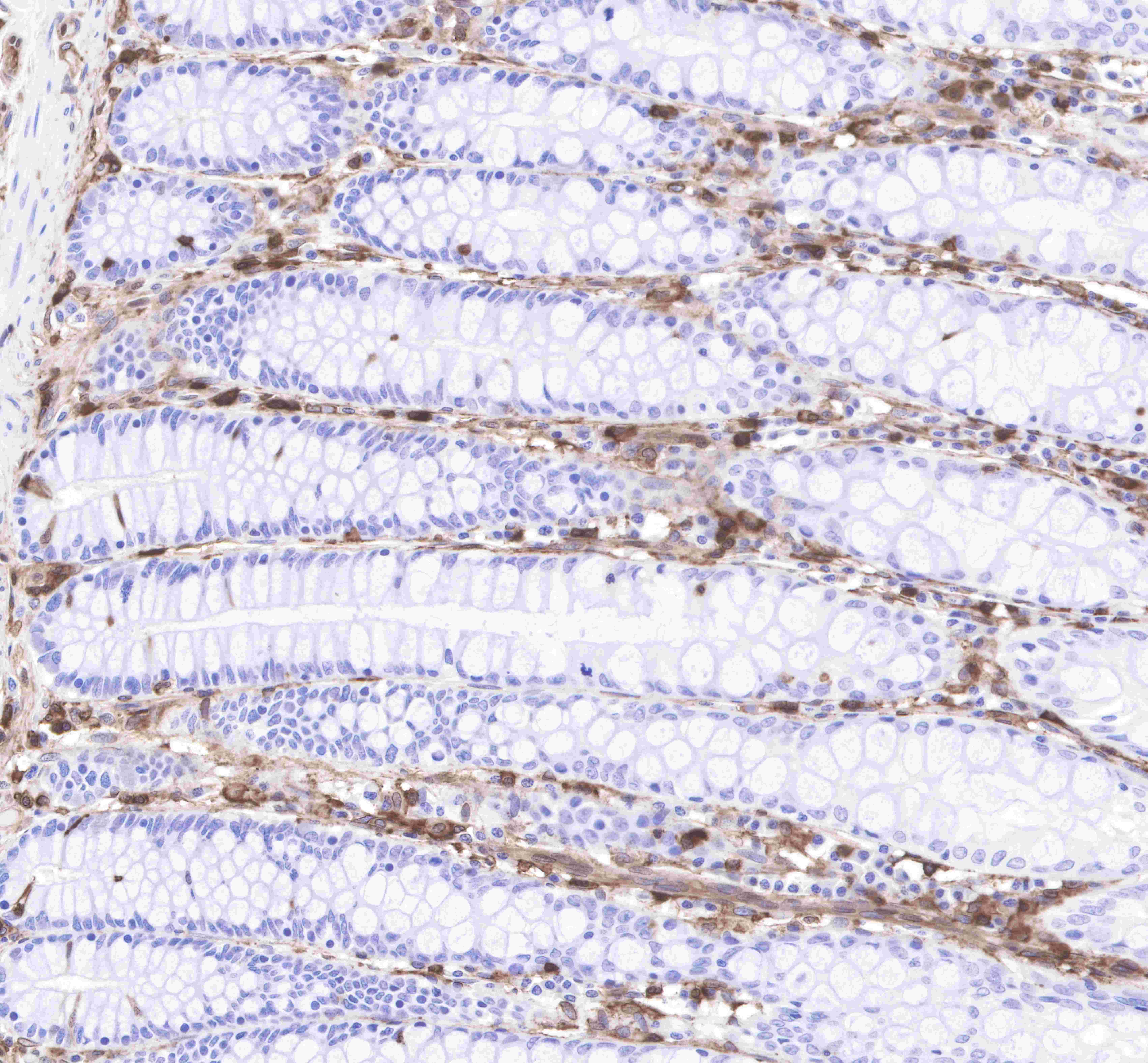
IHC shows positive staining in paraffin-embedded human colon.
Anti-Annexin A1 antibody was used at 1/2000 dilution, followed by a Goat Anti-Rabbit IgG H&L (HRP) ready to use.
Counterstained with hematoxylin.
Heat mediated antigen retrieval with Tris/EDTA buffer pH9.0 was performed before commencing with IHC staining protocol.
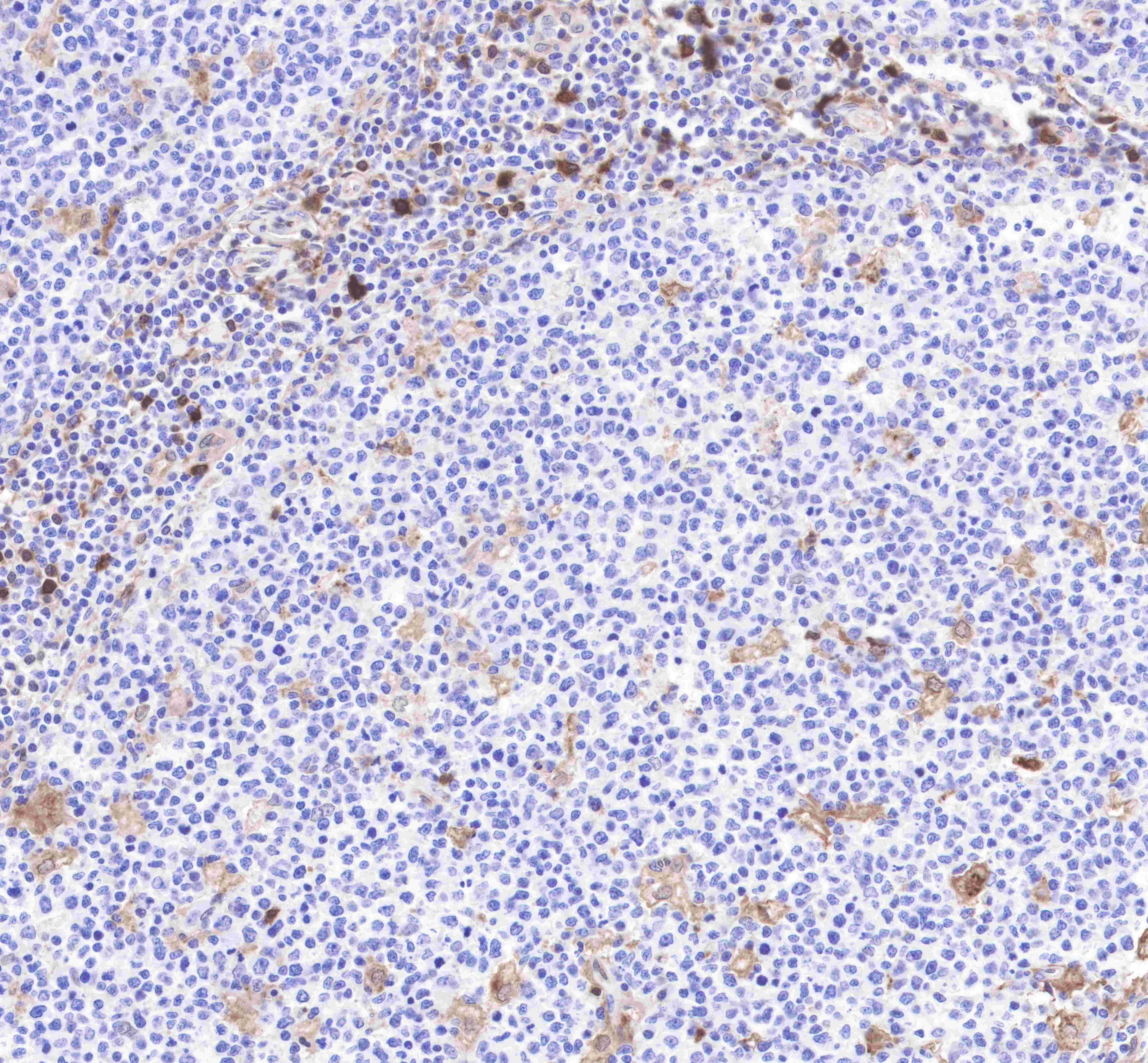
IHC shows positive staining in paraffin-embedded human tonsil.
Anti-Annexin A1 antibody was used at 1/2000 dilution, followed by a Goat Anti-Rabbit IgG H&L (HRP) ready to use.
Counterstained with hematoxylin.
Heat mediated antigen retrieval with Tris/EDTA buffer pH9.0 was performed before commencing with IHC staining protocol.
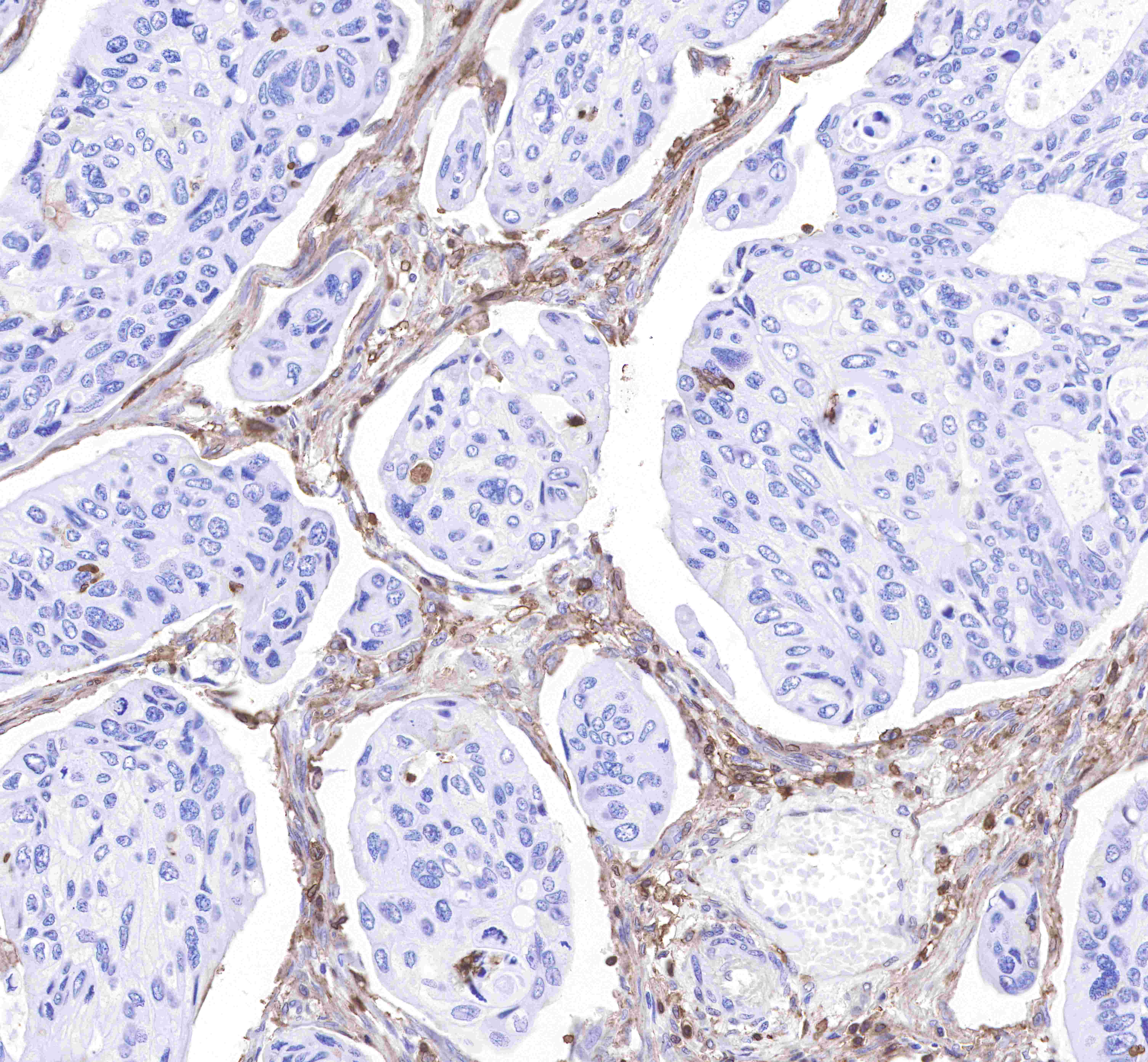
IHC shows positive staining in paraffin-embedded human colon cancer.
Anti-Annexin A1 antibody was used at 1/2000 dilution, followed by a Goat Anti-Rabbit IgG H&L (HRP) ready to use.
Counterstained with hematoxylin.
Heat mediated antigen retrieval with Tris/EDTA buffer pH9.0 was performed before commencing with IHC staining protocol.
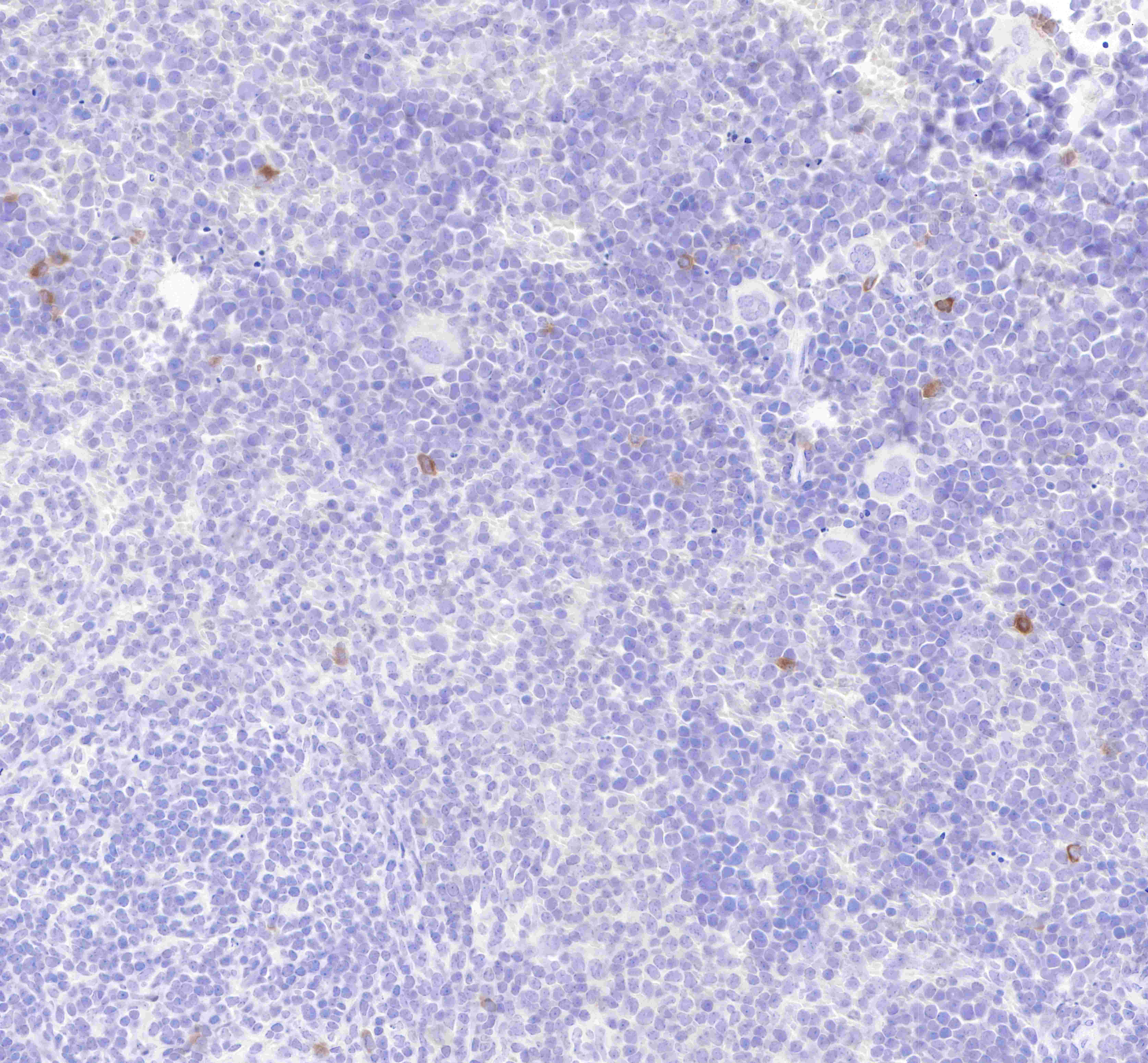
IHC shows positive staining in paraffin-embedded mouse spleen.
Anti-Annexin A1 antibody was used at 1/2000 dilution, followed by a Goat Anti-Rabbit IgG H&L (HRP) ready to use.
Counterstained with hematoxylin.
Heat mediated antigen retrieval with Tris/EDTA buffer pH9.0 was performed before commencing with IHC staining protocol.
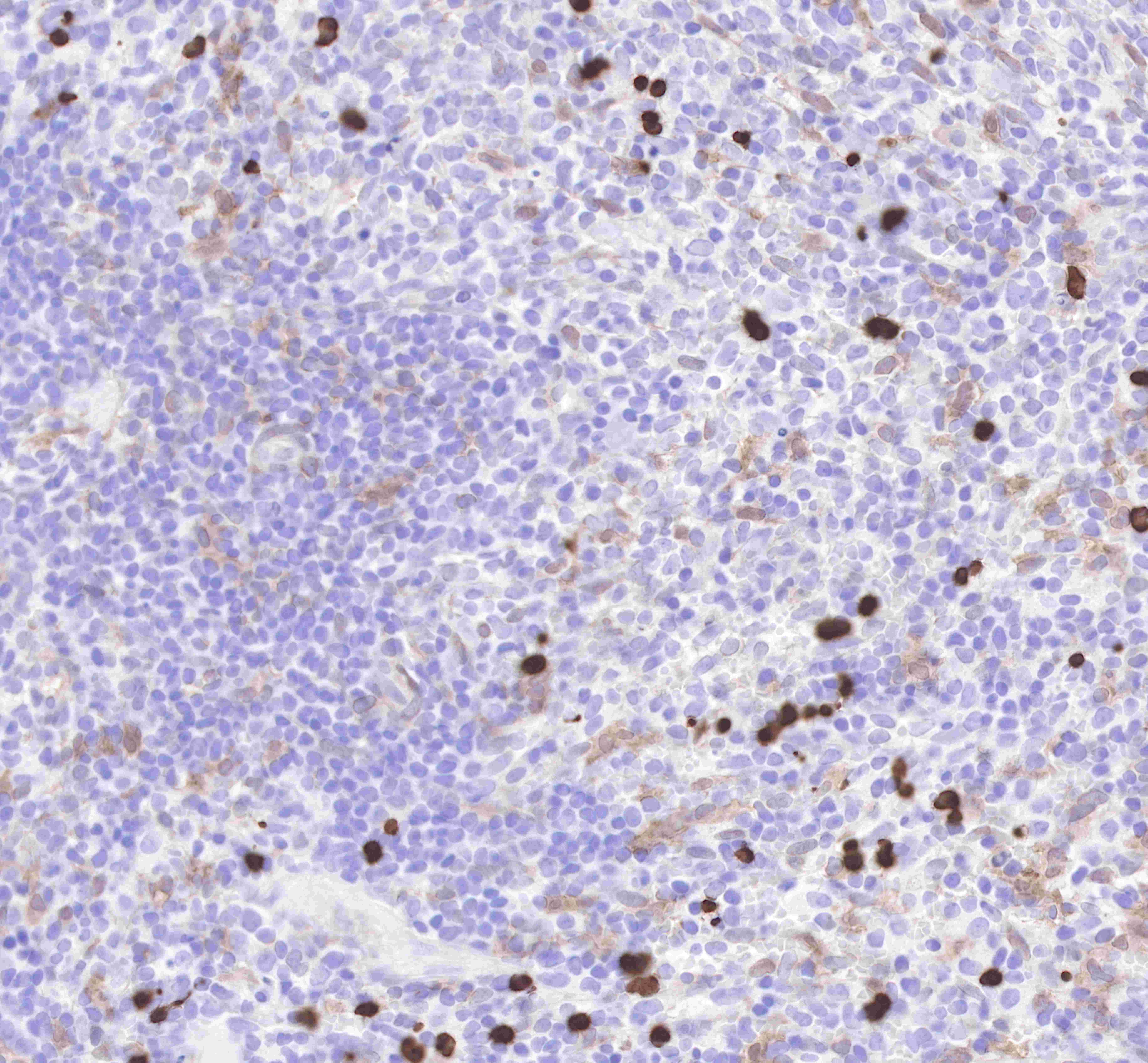
IHC shows positive staining in paraffin-embedded rat spleen.
Anti-Annexin A1 antibody was used at 1/2000 dilution, followed by a Goat Anti-Rabbit IgG H&L (HRP) ready to use.
Counterstained with hematoxylin.
Heat mediated antigen retrieval with Tris/EDTA buffer pH9.0 was performed before commencing with IHC staining protocol.
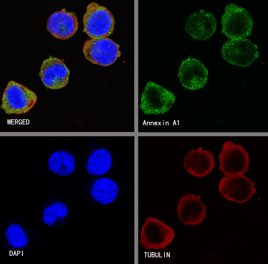
ICC shows positive staining in K562 cells. Anti-Annexin A1 antibody was used at 1/500 dilution (Green) and incubated overnight at 4°C. Goat polyclonal Antibody to rabbit IgG - H&L (Alexa Fluor® 488) was used as secondary antibody at 1/1000 dilution. The cells were fixed with 4% PFA and permeabilized with 0.1% PBS-Triton X-100. Nuclei were counterstained with DAPI (Blue).

Negative control:ICC shows negative staining in 293T cells. Anti-Annexin A1 antibody was used at 1/500 dilution and incubated overnight at 4°C. Goat polyclonal Antibody to rabbit IgG - H&L (Alexa Fluor® 488) was used as secondary antibody at 1/1000 dilution. The cells were fixed with 4% PFA and permeabilized with 0.1% PBS-Triton X-100. Nuclei were counterstained with DAPI (Blue).
 您现在的位置: