
PBS, 40% Glycerol, 0.05% BSA, 0.03% Proclin 300
12 months from date of receipt / reconstitution, -20 °C as supplied
| 应用 | 稀释度 |
|---|---|
| WB | 1:1000 |
| IP | 1:50 |
| ICC | 1:500 |
| ICFCM | 1:50 |
FUS is a multifunctional nuclear protein that is part of the heterogeneous nuclear ribonucleoprotein (hnRNP) complex, involved in processes such as mRNA pre-mRNA splicing and the export of fully processed mRNA to the cytoplasm. FUS belongs to the FET family of RNA-binding proteins and is implicated in various cellular processes, including gene expression regulation, maintenance of genomic integrity, and mRNA/microRNA processing. FUS plays a role in transcription regulation, RNA splicing, RNA transport, DNA repair, and damage response. It binds to nascent pre-mRNAs and acts as a molecular mediator between RNA polymerase II and U1 small nuclear ribonucleoprotein, thereby coupling transcription and splicing. FUS also participates in DNA repair mechanisms by promoting D-loop formation and homologous recombination during DNA double-strand break repair. The abnormal aggregation of FUS is closely related to the occurrence and development of neurodegenerative diseases, such as Amyotrophic Lateral Sclerosis (ALS) and Frontotemporal Dementia (FTD). Under normal conditions, FUS is located in the cell nucleus, but mutations in the gene can cause FUS to be mislocalized to the cytoplasm, where it forms inclusions, which are characteristic of certain neurodegenerative diseases.
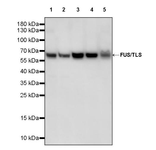
WB result of FUS/TLS Rabbit pAb
Primary antibody: FUS/TLS Rabbit pAb at 1/1000 dilution
Lane 1: HepG2 whole cell lysate 20 µg
Lane 2: K562 whole cell lysate 20 µg
Lane 3: THP-1 whole cell lysate 20 µg
Lane 4: SH-SY5Y whole cell lysate 20 µg
Lane 5: Caco-2 whole cell lysate 20 µg
Secondary antibody: Goat Anti-rabbit IgG, (H+L), HRP conjugated at 1/10000 dilution
Predicted MW: 53 kDa
Observed MW: 65 kDa
WB result of FUS/TLS Rabbit pAb
Primary antibody: FUS/TLS Rabbit pAb at 1/1000 dilution
Lane 1: NIH/3T3 whole cell lysate 20 µg
Lane 2: mouse brain lysate 20 µg
Secondary antibody: Goat Anti-rabbit IgG, (H+L), HRP conjugated at 1/10000 dilution
Predicted MW: 53 kDa
Observed MW: 65 kDa
WB result of FUS/TLS Rabbit pAb
Primary antibody: FUS/TLS Rabbit pAb at 1/1000 dilution
Lane 1: C6 whole cell lysate 20 µg
Lane 2: rat brain lysate 20 µg
Secondary antibody: Goat Anti-rabbit IgG, (H+L), HRP conjugated at 1/10000 dilution
Predicted MW: 53 kDa
Observed MW: 65 kDa
Flow cytometric analysis of HepG2 cells labelling FUS/TLS antibody at 1/50 (1 μg) dilution/ (Red) compared with a Rabbit monoclonal IgG (Black) isotype control and an unlabelled control (cells without incubation with primary antibody and secondary antibody) (Blue). Goat Anti-Rabbit IgG Alexa Fluor® 488 was used as the secondary antibody.
FUS/TLS Rabbit pAb at 1/50 dilution (1 µg) immunoprecipitating FUS/TLS in 0.4 mg K562 whole cell lysate.
Western blot was performed on the immunoprecipitate using FUS/TLS Rabbit pAb at 1/1000 dilution.
Secondary antibody (HRP) for IP was used at 1/1000 dilution.
Lane 1: K562 whole cell lysate 20 µg (Input)
Lane 2: FUS/TLS Rabbit pAb IP in K562 whole cell lysate
Lane 3: Rabbit monoclonal IgG IP in K562 whole cell lysate
Predicted MW: 53 kDa
Observed MW: 65 kDa
ICC shows positive staining in HepG2 cells. Anti-FUS/TLS antibody was used at 1/500 dilution (Green) and incubated overnight at 4°C. Goat polyclonal Antibody to Rabbit IgG - H&L (Alexa Fluor® 488) was used as secondary antibody at 1/1000 dilution. The cells were fixed with 4% PFA and permeabilized with 0.1% PBS-Triton X-100. Nuclei were counterstained with DAPI (Blue). Counterstain with tubulin (Red).
 您现在的位置: