
12 months from date of receipt / reconstitution, -20 °C as supplied
| 应用 | 稀释度 |
|---|---|
| WB | 1:500-1:1000 |
| IP | 1:100 |
| ICC | 1:1000 |
Histone H3 crotonylation, specifically at lysine 9 (H3K9cr), is a form of post-translational modification that has been identified as a key player in epigenetic regulation, particularly in plants 。This modification is structurally and functionally distinct from the well-studied lysine acetylation and is involved in various biological processes, including chromatin remodeling, metabolism, cell cycle, and cellular organization. In the context of histones, crotonylation can be found on different lysine residues among plant species and is known to overlap with the histone modification H3K9ac in about 95% of the regions, which suggests a potential interplay between these two modifications.
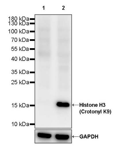
WB result of Histone H3 (Crotonyl K9) Recombinant Rabbit mAb
Primary antibody: Histone H3 (Crotonyl K9) Recombinant Rabbit mAb at 1/1000 dilution
Lane 1: untreated HeLa whole cell lysate 20 µg
Lane 2: HeLa treated with 500 ng/ml TSA for 4 hours whole cell lysate 20 µg
Secondary antibody: Goat Anti-rabbit IgG, (H+L), HRP conjugated at 1/10000 dilution
Predicted MW: 15 kDa
Observed MW: 17 kDa
This blot was developed with high sensitivity substrate
Histone H3 (Crotonyl K9) Rabbit mAb at 1/100 dilution (1 µg) immunoprecipitating Histone H3 (Crotonyl K9) in 0.4 mg HeLa treated with 500 ng/ml TSA for 4 hours whole cell lysate.
Western blot was performed on the immunoprecipitate using Histone H3 (Crotonyl K9) Rabbit mAb at 1/1000 dilution.
Secondary antibody (HRP) for IP was used at 1/1000 dilution.
Lane 1: HeLa treated with 500 ng/ml TSA for 4 hours whole cell lysate 20 µg (Input)
Lane 2: Histone H3 (Crotonyl K9) Rabbit mAb IP in HeLa treated with 500 ng/ml TSA for 4 hours whole cell lysate
Lane 3: Rabbit monoclonal IgG IP in HeLa treated with 500 ng/ml TSA for 4 hours whole cell lysate
Predicted MW: 15 kDa
Observed MW: 17 kDa
This blot was developed with high sensitivity substrate
ICC analysis of HeLa cells treated with TSA (500ng/ml,4h) (top panel) and HeLa cells untreated with TSA (500ng/ml,4h) (below panel). Anti-Histone H3 (Crotonyl K9) antibody was used at 1/1000 dilution (Green) and incubated overnight at 4°C. Goat polyclonal Antibody to Rabbit IgG - H&L (Alexa Fluor® 488) was used as secondary antibody at 1/1000 dilution. The cells were fixed with 4% PFA and permeabilized with 0.1% PBS-Triton X-100. Nuclei were counterstained with DAPI (Blue). Counterstain with tubulin (Red).
 您现在的位置: