
PBS, 40% Glycerol, 0.05% BSA, 0.03% Proclin 300
12 months from date of receipt / reconstitution, -20 °C as supplied
| 应用 | 稀释度 |
|---|---|
| WB | 1:1000 |
| IP | 1:50 |
| ICC | 1:1000 |
Microphthalmia-associated transcription factor (MiTF) is a protein that plays a crucial role in the development and function of several cell types, including melanocytes, osteoclasts, and certain types of neurons. It is a member of the bHLH-LZ (basic helix-loop-helix-leucine zipper) family of transcription factors, which are characterized by their ability to bind DNA and regulate gene expression through dimerization. MiTF is involved in the regulation of pigmentation, particularly in the melanocytes of the skin, hair, and eyes. It activates the expression of genes involved in melanin synthesis, which is essential for the coloration of various tissues and organs. Mutations in the MITF gene can lead to conditions such as Waardenburg syndrome, which is characterized by hearing loss and pigmentation abnormalities. In addition to its role in pigmentation, MiTF is also implicated in the development of the immune system, particularly in the differentiation of T cells. It has been shown to regulate the expression of genes involved in T cell development and function. Furthermore, MiTF is involved in the regulation of bone homeostasis. It is expressed in osteoclasts and is thought to play a role in bone resorption and remodeling. Disruptions in MiTF function can lead to bone disorders, such as osteopetrosis, a condition characterized by increased bone density and reduced bone marrow space.
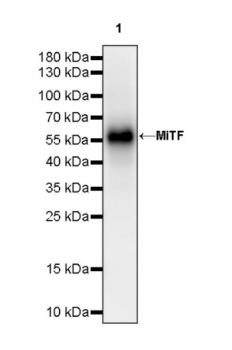
WB result of MiTF Recombinant Rabbit mAb
Primary antibody: MiTF Recombinant Rabbit mAb at 1/1000 dilution
Lane 1: SK-MEL-28 whole cell lysate 20 µg
Secondary antibody: Goat Anti-rabbit IgG, (H+L), HRP conjugated at 1/10000 dilution
Predicted MW: 58 kDa
Observed MW: 58 kDa
This blot was developed with high sensitivity substrate
WB result of MiTF Recombinant Rabbit mAb
Primary antibody: MiTF Recombinant Rabbit mAb at 1/1000 dilution
Lane 1: B16-F0 whole cell lysate 20 µg
Secondary antibody: Goat Anti-rabbit IgG, (H+L), HRP conjugated at 1/10000 dilution
Predicted MW: 58 kDa
Observed MW: 58 kDa
This blot was developed with high sensitivity substrate
MiTF Rabbit mAb at 1/50 dilution (1 µg) immunoprecipitating MiTF in 0.4 mg SK-MEL-28 whole cell lysate.
Western blot was performed on the immunoprecipitate using MiTF Rabbit mAb at 1/1000 dilution.
Secondary antibody (HRP) for IP was used at 1/1000 dilution.
Lane 1: SK-MEL-28 whole cell lysate 20 µg (Input)
Lane 2: MiTF Rabbit mAb IP in SK-MEL-28 whole cell lysate
Lane 3: Rabbit monoclonal IgG IP in SK-MEL-28 whole cell lysate
Predicted MW: 58 kDa
Observed MW: 58 kDa
ICC shows positive staining in SK-MEL-28 cells (top panel) and negative staining in HCT116 cells (below panel). Anti-MiTF antibody was used at 1/500 dilution (Green) and incubated overnight at 4°C. Goat polyclonal Antibody to Rabbit IgG - H&L (Alexa Fluor® 488) was used as secondary antibody at 1/1000 dilution. The cells were fixed with 100% ice-cold methanol and permeabilized with 0.1% PBS-Triton X-100. Nuclei were counterstained with DAPI (Blue). Counterstain with tubulin (Red).
 您现在的位置: