
PBS, 40% Glycerol, 0.05% BSA, 0.03% Proclin 300
12 months from date of receipt / reconstitution, -20 °C as supplied
| 应用 | 稀释度 |
|---|---|
| WB | 1:1000 |
| ICC | 1:500 |
| FCM | 1:50 |
NG2 Chondroitin Sulfate Proteoglycan, also known as neuron-glial antigen 2 (NG2) or chondroitin sulfate proteoglycan 4 (CSPG4), is a cell surface molecule that is involved in a variety of cellular processes. It is a type I transmembrane core proteoglycan that is crucial for cell survival, migration, and angiogenesis. The regulation of NG2 expression is influenced by factors such as inflammation and hypoxia and is mediated by methyltransferases, transcription factors including Sp1, Pax3, Egr-1, and the microRNA miR129-2. These regulatory elements are essential in determining cellular processes mediated by NG2, such as glial scar formation in the central nervous system (CNS) or tumor growth and metastasis. NG2 has been implicated in the progression of several tumor types, with elevated NG2 expression found predominantly in glioblastoma, correlating with poor prognosis due to increased chemo- and radioresistance of tumor cells. It is also a key intermediate of tumor cells with extracellular matrix molecules, determining metastatic formation in soft-tissue sarcoma and melanoma patients. In addition to its role in tumorigenesis, NG2 is expressed in certain benign cell types, such as NG2-glia in the CNS, mesenchymal stem cells, osteoblasts, melanocytes, smooth muscle cells, and macrophages. It is also a typical marker for pericytes, which contribute to the stabilization of microvessels, regulation of capillary blood flow, and angiogenesis. In the context of inflammation, NG2 expression is induced in activated microglial cells, and it mediates the induction of inducible nitric oxide synthase (iNOS) and inflammatory cytokine expression, but not chemokine expression in activated microglia. This suggests that NG2 plays a role in the inflammatory response of these cells.
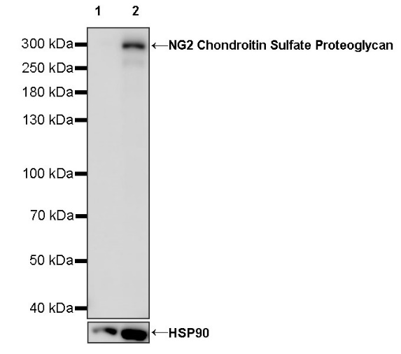
WB result of NG2 Chondroitin Sulfate Proteoglycan Recombinant Rabbit mAb
Primary antibody: NG2 Chondroitin Sulfate Proteoglycan Recombinant Rabbit mAb at 1/1000 dilution
Lane 1: mouse liver lysate 20 µg
Lane 2: mouse brain lysate 20 µg
Negative control: mouse liver lysate
Secondary antibody: Goat Anti-rabbit IgG, (H+L), HRP conjugated at 1/10000 dilution
Predicted MW: 252 kDa
Observed MW: 300 kDa
WB result of NG2 Chondroitin Sulfate Proteoglycan Recombinant Rabbit mAb
Primary antibody: NG2 Chondroitin Sulfate Proteoglycan Recombinant Rabbit mAb at 1/1000 dilution
Lane 1: rat brain lysate 20 µg
Secondary antibody: Goat Anti-rabbit IgG, (H+L), HRP conjugated at 1/10000 dilution
Predicted MW: 252 kDa
Observed MW: 300 kDa
Flow cytometric analysis of MCF7 (Human breast adenocarcinoma epithelial cell, left) / A375 (Human malignant melanoma epithelial cell, right) labelling NG2 Chondroitin Sulfate Proteoglycan antibody at 1/50 dilution (1 μg) / (Red) compared with a Rabbit monoclonal IgG (Black) isotype control and an unlabelled control (cells without incubation with primary antibody and secondary antibody) (Blue). Goat Anti - Rabbit IgG Alexa Fluor® 488 was used as the secondary antibody.
Negative control: MCF7
ICC shows positive staining in A375 cells (top panel) and negative staining in MCF7 cells (below panel). Anti- NG2 Chondroitin Sulfate Proteoglycan antibody was used at 1/500 dilution (Green) and incubated overnight at 4°C. Goat polyclonal Antibody to Rabbit IgG - H&L (Alexa Fluor® 488) was used as secondary antibody at 1/1000 dilution. The cells were fixed with 4% PFA and permeabilized with 0.1% PBS-Triton X-100. Nuclei were counterstained with DAPI (Blue). Counterstain with tubulin (Red).
 您现在的位置: