
PBS, 40% Glycerol, 0.05% BSA, 0.03% Proclin 300
12 months from date of receipt / reconstitution, -20 °C as supplied
| 应用 | 稀释度 |
|---|---|
| WB | 1:1000 |
| IP | 1:50 |
| IHC-P | 1:250-1:500 |
| ICC | 1:500 |
| ICFCM | 1:500 |
H2AFY is a protein that is encoded by the H2AFY gene in humans. It is a member of the histone H2A family and is involved in the regulation of chromatin structure and gene expression. H2AFY is one of the histone variants that are enriched on the inactive X chromosome in female mammals, contributing to the process of dosage compensation where one of the two X chromosomes is inactivated to balance the gene expression levels between males and females. Alterations in the expression of H2AFY have been observed in various types of cancer. It has been suggested that H2AFY may play a role in tumorigenesis and could be a potential therapeutic target for cancer treatment. H2AFY has been linked to the regulation of the cell cycle, with its expression levels varying throughout the different stages of the cell cycle. H2AFY can influence gene expression by affecting the chromatin structure, which in turn affects the binding of transcription factors and the transcriptional machinery. H2AFY may also be involved in the cellular response to DNA damage, potentially playing a role in the repair mechanisms that maintain genomic integrity.
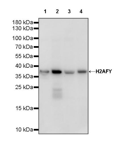
WB result of H2AFY Recombinant Rabbit mAb
Primary antibody: H2AFY Recombinant Rabbit mAb at 1/1000 dilution
Lane 1: HeLa whole cell lysate 20 µg
Lane 2: HepG2 whole cell lysate 20 µg
Lane 3: MCF7 whole cell lysate 20 µg
Lane 4: 293T whole cell lysate 20 µg
Secondary antibody: Goat Anti-rabbit IgG, (H+L), HRP conjugated at 1/10000 dilution
Predicted MW: 39 kDa
Observed MW: 38 kDa
WB result of H2AFY Recombinant Rabbit mAb
Primary antibody: H2AFY Recombinant Rabbit mAb at 1/1000 dilution
Lane 1: NIH/3T3 whole cell lysate 20 µg
Secondary antibody: Goat Anti-rabbit IgG, (H+L), HRP conjugated at 1/10000 dilution
Predicted MW: 39 kDa
Observed MW: 38 kDa
WB result of H2AFY Recombinant Rabbit mAb
Primary antibody: H2AFY Recombinant Rabbit mAb at 1/1000 dilution
Lane 1: C6 whole cell lysate 20 µg
Secondary antibody: Goat Anti-rabbit IgG, (H+L), HRP conjugated at 1/10000 dilution
Predicted MW: 39 kDa
Observed MW: 38 kDa
Flow cytometric analysis of 4% PFA fixed 90% methanol permeabilized HeLa (Human cervix adenocarcinoma epithelial cell) labelling H2AFY antibody at 1/500 dilution (0.1 μg) / (Red) compared with a Rabbit monoclonal IgG (Black) isotype control and an unlabelled control (cells without incubation with primary antibody and secondary antibody) (Blue). Goat Anti - Rabbit IgG Alexa Fluor® 488 was used as the secondary antibody.
H2AFY Rabbit mAb at 1/50 dilution (1 µg) immunoprecipitating H2AFY in 0.4 mg 293T whole cell lysate.
Western blot was performed on the immunoprecipitate using H2AFY Rabbit mAb at 1/1000 dilution.
Secondary antibody (HRP) for IP was used at 1/1000 dilution.
Lane 1: 293T whole cell lysate 20 µg (Input)
Lane 2: H2AFY Rabbit mAb IP in 293T whole cell lysate
Lane 3: Rabbit monoclonal IgG IP in 293T whole cell lysate
Predicted MW: 39 kDa
Observed MW: 38 kDa
This blot was developed with high sensitivity substrate
IHC shows positive staining in paraffin-embedded human colon. Anti-H2AFY antibody was used at 1/250 dilution, followed by a HRP Polymer for Mouse & Rabbit IgG (ready to use). Counterstained with hematoxylin. Heat mediated antigen retrieval with Tris/EDTA buffer pH9.0 was performed before commencing with IHC staining protocol.
IHC shows positive staining in paraffin-embedded human liver. Anti-H2AFY antibody was used at 1/250 dilution, followed by a HRP Polymer for Mouse & Rabbit IgG (ready to use). Counterstained with hematoxylin. Heat mediated antigen retrieval with Tris/EDTA buffer pH9.0 was performed before commencing with IHC staining protocol.
IHC shows positive staining in paraffin-embedded human thyroid cancer. Anti-H2AFY antibody was used at 1/250 dilution, followed by a HRP Polymer for Mouse & Rabbit IgG (ready to use). Counterstained with hematoxylin. Heat mediated antigen retrieval with Tris/EDTA buffer pH9.0 was performed before commencing with IHC staining protocol.
IHC shows positive staining in paraffin-embedded human ovarian cancer. Anti-H2AFY antibody was used at 1/250 dilution, followed by a HRP Polymer for Mouse & Rabbit IgG (ready to use). Counterstained with hematoxylin. Heat mediated antigen retrieval with Tris/EDTA buffer pH9.0 was performed before commencing with IHC staining protocol.
IHC shows positive staining in paraffin-embedded mouse cerebral cortex. Anti-H2AFY antibody was used at 1/250 dilution, followed by a HRP Polymer for Mouse & Rabbit IgG (ready to use). Counterstained with hematoxylin. Heat mediated antigen retrieval with Tris/EDTA buffer pH9.0 was performed before commencing with IHC staining protocol.
IHC shows positive staining in paraffin-embedded rat kidney. Anti-H2AFY antibody was used at 1/250 dilution, followed by a HRP Polymer for Mouse & Rabbit IgG (ready to use). Counterstained with hematoxylin. Heat mediated antigen retrieval with Tris/EDTA buffer pH9.0 was performed before commencing with IHC staining protocol.
ICC shows positive staining in HeLa cells. Anti-H2AFY antibody was used at 1/500 dilution (Green) and incubated overnight at 4°C. Goat polyclonal Antibody to Rabbit IgG - H&L (Alexa Fluor® 488) was used as secondary antibody at 1/1000 dilution. The cells were fixed with 100% ice-cold methanol and permeabilized with 0.1% PBS-Triton X-100. Nuclei were counterstained with DAPI (Blue). Counterstain with tubulin (Red).
 您现在的位置: