
PBS, 40% Glycerol, 0.05% BSA, 0.03% Proclin 300
12 months from date of receipt / reconstitution, -20 °C as supplied
| 应用 | 稀释度 |
|---|---|
| WB | 1:1000 |
| IP | 1:50 |
| IHC-P | 1:250 |
| ICC | 1:100 |
| ICFCM | 1:500 |
JNK1, also known as c-Jun N-terminal kinase 1, is a member of the mitogen-activated protein kinase (MAPK) family of enzymes that play essential roles in cellular signaling pathways. JNK1 is primarily involved in the cellular response to stress. JNK1 is activated by a complex signaling cascade involving upstream kinases, such as MEKK1, MLK3, and ASK1. Activation typically occurs in response to stress stimuli, which trigger the assembly of a multi-protein complex known as the MAPKKK-MAPKK-MAPK module. This complex leads to the phosphorylation and activation of JNK1, which then phosphorylates its downstream targets. Upon activation, it phosphorylates specific substrates, including transcription factors such as c-Jun, ATF2, and p53, leading to changes in gene expression that help cells adapt to stress. JNK1 plays a dual role in regulating cell death (apoptosis) and survival. Depending on the cellular context and stimuli, JNK1 can either promote or inhibit apoptosis. JNK1 is also involved in inflammatory and immune responses. It can regulate the production of cytokines, chemokines, and other inflammatory mediators, as well as modulate the activation and function of immune cells.
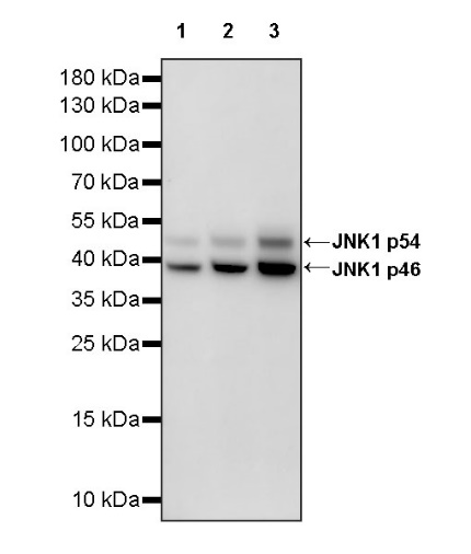
WB result of JNK1 Recombinant Rabbit mAb
Primary antibody: JNK1 Recombinant Rabbit mAb at 1/1000 dilution
Lane 1: A431 whole cell lysate 20 µg
Lane 2: HeLa whole cell lysate 20 µg
Lane 3: Jurkat whole cell lysate 20 µg
Secondary antibody: Goat Anti-rabbit IgG, (H+L), HRP conjugated at 1/10000 dilution
Predicted MW: 44 kDa
Observed MW: 38, 50 kDa
WB result of JNK1 Recombinant Rabbit mAb
Primary antibody: JNK1 Recombinant Rabbit mAb at 1/1000 dilution
Lane 1: NIH/3T3 whole cell lysate 20 µg
Lane 2: Neuro-2a whole cell lysate 20 µg
Secondary antibody: Goat Anti-rabbit IgG, (H+L), HRP conjugated at 1/10000 dilution
Predicted MW: 44 kDa
Observed MW: 38, 50 kDa
WB result of JNK1 Recombinant Rabbit mAb
Primary antibody: JNK1 Recombinant Rabbit mAb at 1/1000 dilution
Lane 1: PC-12 whole cell lysate 20 µg
Lane 2: C6 whole cell lysate 20 µg
Secondary antibody: Goat Anti-rabbit IgG, (H+L), HRP conjugated at 1/10000 dilution
Predicted MW: 44 kDa
Observed MW: 38, 50 kDa
Flow cytometric analysis of 4% PFA fixed 90% methanol permeabilized HeLa (Human cervix adenocarcinoma epithelial cell) labelling JNK1 antibody at 1/500 dilution (0.1 μg) / (Red) compared with a Rabbit monoclonal IgG (Black) isotype control and an unlabelled control (cells without incubation with primary antibody and secondary antibody) (Blue). Goat Anti - Rabbit IgG Alexa Fluor® 488 was used as the secondary antibody.
JNK1 Rabbit mAb at 1/50 dilution (1 µg) immunoprecipitating JNK1 in 0.4 mg Neuro-2a whole cell lysate.
Western blot was performed on the immunoprecipitate using JNK1 Rabbit mAb at 1/1000 dilution.
Secondary antibody (HRP) for IP was used at 1/1000 dilution.
Lane 1: Neuro-2a whole cell lysate 20 µg (Input)
Lane 2: JNK1 Rabbit mAb IP in Neuro-2a whole cell lysate
Lane 3: Rabbit monoclonal IgG IP in Neuro-2a whole cell lysate
Predicted MW: 44 kDa
Observed MW: 38, 50 kDa
IHC shows positive staining in paraffin-embedded human ovarian carcinoma. Anti-JNK1 antibody was used at 1/250 dilution, followed by a HRP Polymer for Mouse & Rabbit IgG (ready to use). Counterstained with hematoxylin. Heat mediated antigen retrieval with Tris/EDTA buffer pH9.0 was performed before commencing with IHC staining protocol.
IHC shows positive staining in paraffin-embedded human prostatic carcinoma. Anti-JNK1 antibody was used at 1/250 dilution, followed by a HRP Polymer for Mouse & Rabbit IgG (ready to use). Counterstained with hematoxylin. Heat mediated antigen retrieval with Tris/EDTA buffer pH9.0 was performed before commencing with IHC staining protocol.
IHC shows positive staining in paraffin-embedded mouse cerebral cortex. Anti-JNK1 antibody was used at 1/250 dilution, followed by a HRP Polymer for Mouse & Rabbit IgG (ready to use). Counterstained with hematoxylin. Heat mediated antigen retrieval with Tris/EDTA buffer pH9.0 was performed before commencing with IHC staining protocol.
IHC shows positive staining in paraffin-embedded mouse testis. Anti-JNK1 antibody was used at 1/250 dilution, followed by a HRP Polymer for Mouse & Rabbit IgG (ready to use). Counterstained with hematoxylin. Heat mediated antigen retrieval with Tris/EDTA buffer pH9.0 was performed before commencing with IHC staining protocol.
IHC shows positive staining in paraffin-embedded rat testis. Anti-JNK1 antibody was used at 1/250 dilution, followed by a HRP Polymer for Mouse & Rabbit IgG (ready to use). Counterstained with hematoxylin. Heat mediated antigen retrieval with Tris/EDTA buffer pH9.0 was performed before commencing with IHC staining protocol.
ICC shows positive staining in HeLa cells. Anti-JNK1 antibody was used at 1/100 dilution (Green) and incubated overnight at 4°C. Goat polyclonal Antibody to Rabbit IgG - H&L (Alexa Fluor® 488) was used as secondary antibody at 1/1000 dilution. The cells were fixed with 100% ice-cold methanol and permeabilized with 0.1% PBS-Triton X-100. Nuclei were counterstained with DAPI (Blue). Counterstain with tubulin (Red).
 您现在的位置: