
12 months from date of receipt / reconstitution, -20 °C as supplied
| 应用 | 稀释度 |
|---|---|
| WB | 1:1000 |
| IHC-P | 1:100 |
| IP | 1:50 |
FGL1 (Fibrinogen-Like Protein 1) is a protein that belongs to the fibrinogen-related protein superfamily. This family of proteins shares structural similarities with fibrinogen, a major component of blood clots, but they have diverse functions beyond coagulation. FGL1 is expressed in various tissues and cell types, including immune cells such as macrophages and dendritic cells. It has been implicated in several biological processes, particularly in the immune system. One of its key roles is in the regulation of innate and adaptive immune responses. FGL1 has been shown to interact with several immune receptors and molecules, modulating their signaling pathways. For example, it can bind to certain inhibitory receptors on immune cells, such as TIGIT (T-cell immunoreceptor with Ig and ITIM domains), and mediate immune suppression. This interaction is thought to contribute to the regulation of immune tolerance and the prevention of autoimmune diseases. In addition to its role in immune regulation, FGL1 has also been linked to cancer biology. Studies have shown that FGL1 expression is altered in some cancer types, and it may play a role in tumor growth, metastasis, and immune evasion.
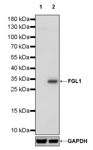
WB result of FGL1 Recombinant Rabbit mAb
Primary antibody: FGL1 Recombinant Rabbit mAb at 1/1000 dilution
Lane 1: HCT 116 whole cell lysate 20 µg
Lane 2: HepG2 whole cell lysate 20 µg
Negative control: HCT 116 whole cell lysate
Secondary antibody: Goat Anti-rabbit IgG, (H+L), HRP conjugated at 1/10000 dilution
Predicted MW: 36 kDa
Observed MW: 34 kDa
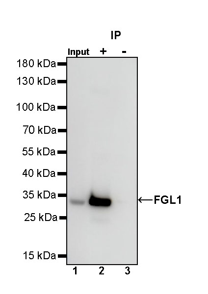
FGL1 Rabbit mAb at 1/50 dilution (1 µg) immunoprecipitating FGL1 in 0.4 mg HepG2 whole cell lysate.
Western blot was performed on the immunoprecipitate using FGL1 Rabbit mAb at 1/1000 dilution.
Secondary antibody (HRP) for IP was used at 1/1000 dilution.
Lane 1: HepG2 whole cell lysate 20 µg (Input)
Lane 2: FGL1 Rabbit mAb IP in HepG2 whole cell lysate
Lane 3: Rabbit monoclonal IgG IP in HepG2 whole cell lysate
Predicted MW: 36 kDa
Observed MW: 34 kDa
IHC shows positive staining in paraffin-embedded human liver. Anti-FGL1 antibody was used at 1/100 dilution, followed by a HRP Polymer for Mouse & Rabbit IgG (ready to use). Counterstained with hematoxylin. Heat mediated antigen retrieval with Tris/EDTA buffer pH9.0 was performed before commencing with IHC staining protocol.
Negative control: IHC shows negative staining in paraffin-embedded human cerebral cortex. Anti-FGL1 antibody was used at 1/100 dilution, followed by a HRP Polymer for Mouse & Rabbit IgG (ready to use). Counterstained with hematoxylin. Heat mediated antigen retrieval with Tris/EDTA buffer pH9.0 was performed before commencing with IHC staining protocol.
IHC shows positive staining in paraffin-embedded human hepatocellular carcinoma. Anti-FGL1 antibody was used at 1/100 dilution, followed by a HRP Polymer for Mouse & Rabbit IgG (ready to use). Counterstained with hematoxylin. Heat mediated antigen retrieval with Tris/EDTA buffer pH9.0 was performed before commencing with IHC staining protocol.
 您现在的位置: