
PBS, 40% Glycerol, 0.05% BSA, 0.03% Proclin 300
12 months from date of receipt / reconstitution, -20 °C as supplied
| 应用 | 稀释度 |
|---|---|
| WB | 1:1000 |
| IP | 1:50 |
| IHC-P | 1:1000 |
| ICC | 1:500 |
| ICFCM | 1:500 |
NaK-ATPase, also known as the sodium-potassium adenosine triphosphatase (Na+/K+-ATPase) or simply the sodium pump, is a crucial transmembrane protein found in the plasma membrane of animal cells. Utilizing the energy released from ATP hydrolysis, Na+/K+-ATPase actively transports Na+ ions out of the cell against their concentration gradient while simultaneously transporting K+ ions into the cell. This ion transport activity is essential for maintaining the electrochemical gradients across the cell membrane, contributing to the stability of the intracellular and extracellular environments. In addition to its role as an ion pump, Na+/K+-ATPase has signaling functions. It can serve as a receptor for certain hormones and drugs, participating in intracellular signaling cascades.
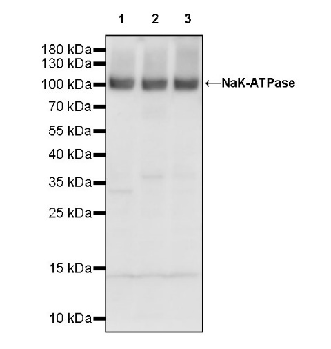
WB result of NaK-ATPase Recombinant Rabbit mAb
Primary antibody: NaK-ATPase Recombinant Rabbit mAb at 1/1000 dilution
Lane 1: unboiled HeLa whole cell lysate 20 µg
Lane 2: unboiled MCF-7 whole cell lysate 20 µg
Lane 3: unboiled HEK-293 whole cell lysate 20 µg
Secondary antibody: Goat Anti-rabbit IgG, (H+L), HRP conjugated at 1/10000 dilution
Predicted MW: 113 kDa
Observed MW: 110 kDa
WB result of NaK-ATPase Recombinant Rabbit mAb
Primary antibody: NaK-ATPase Recombinant Rabbit mAb at 1/1000 dilution
Lane 1: unboiled C2C12 whole cell lysate 20 µg
Secondary antibody: Goat Anti-rabbit IgG, (H+L), HRP conjugated at 1/10000 dilution
Predicted MW: 113 kDa
Observed MW: 110 kDa
WB result of NaK-ATPase Recombinant Rabbit mAb
Primary antibody: NaK-ATPase Recombinant Rabbit mAb at 1/1000 dilution
Lane 1: unboiled C6 whole cell lysate 20 µg
Secondary antibody: Goat Anti-rabbit IgG, (H+L), HRP conjugated at 1/10000 dilution
Predicted MW: 113 kDa
Observed MW: 110 kDa
Flow cytometric analysis of 4% PFA fixed 90% methanol permeabilized MCF7 (Human breast adenocarcinoma epithelial cell) labelling NaK-ATPase antibody at 1/500 dilution (0.1 μg)/ (Red) compared with a Rabbit monoclonal IgG (Black) isotype control and an unlabelled control (cells without incubation with primary antibody and secondary antibody) (Blue). Goat Anti - Rabbit IgG Alexa Fluor® 488 was used as the secondary antibody.
NaK-ATPase Rabbit mAb at 1/50 dilution (1 µg) immunoprecipitating NaK-ATPase in 0.4 mg unboiled HeLa whole cell lysate.
Western blot was performed on the immunoprecipitate using NaK-ATPase Rabbit mAb at 1/1000 dilution.
Secondary antibody (HRP) for IP was used at 1/1000 dilution.
Lane 1: unboiled HeLa whole cell lysate 20 µg (Input)
Lane 2: NaK-ATPase Rabbit mAb IP in unboiled HeLa whole cell lysate
Lane 3: Rabbit monoclonal IgG IP in unboiled HeLa whole cell lysate
Predicted MW: 113 kDa
Observed MW: 113 kDa
IHC shows positive staining in paraffin-embedded human kidney. Anti- NaK-ATPase antibody was used at 1/1000 dilution, followed by a HRP Polymer for Mouse & Rabbit IgG (ready to use). Counterstained with hematoxylin. Heat mediated antigen retrieval with Tris/EDTA buffer pH9.0 was performed before commencing with IHC staining protocol.
IHC shows positive staining in paraffin-embedded human liver. Anti- NaK-ATPase antibody was used at 1/1000 dilution, followed by a HRP Polymer for Mouse & Rabbit IgG (ready to use). Counterstained with hematoxylin. Heat mediated antigen retrieval with Tris/EDTA buffer pH9.0 was performed before commencing with IHC staining protocol.
Negative control: IHC shows negative staining in paraffin-embedded human spleen. Anti- NaK-ATPase antibody was used at 1/1000 dilution, followed by a HRP Polymer for Mouse & Rabbit IgG (ready to use). Counterstained with hematoxylin. Heat mediated antigen retrieval with Tris/EDTA buffer pH9.0 was performed before commencing with IHC staining protocol.
IHC shows positive staining in paraffin-embedded human hepatocellular carcinoma. Anti- NaK-ATPase antibody was used at 1/1000 dilution, followed by a HRP Polymer for Mouse & Rabbit IgG (ready to use). Counterstained with hematoxylin. Heat mediated antigen retrieval with Tris/EDTA buffer pH9.0 was performed before commencing with IHC staining protocol.
IHC shows positive staining in paraffin-embedded human colon cancer. Anti- NaK-ATPase antibody was used at 1/1000 dilution, followed by a HRP Polymer for Mouse & Rabbit IgG (ready to use). Counterstained with hematoxylin. Heat mediated antigen retrieval with Tris/EDTA buffer pH9.0 was performed before commencing with IHC staining protocol.
ICC shows positive staining in MCF-7 cells. Anti-NaK-ATPase antibody was used at 1/500 dilution (Green) and incubated overnight at 4°C. Goat polyclonal Antibody to Rabbit IgG - H&L (Alexa Fluor® 488) was used as secondary antibody at 1/1000 dilution. The cells were fixed with 100% ice-cold methanol and permeabilized with 0.1% PBS-Triton X-100. Nuclei were counterstained with DAPI (Blue). Counterstain with tubulin (Red).
 您现在的位置: