
PBS, 40% Glycerol, 0.05% BSA, 0.03% Proclin 300
12 months from date of receipt / reconstitution, -20 °C as supplied
| 应用 | 稀释度 |
|---|---|
| WB | 1:1000 |
| IP | 1:50 |
| IHC-P | 1:500 |
| ICC | 1:500 |
| ICFCM | 1:50 |
LKB1 is a serine/threonine kinase that belongs to the AMPK-related kinase family. LKB1 functions as a heterotrimeric complex with STRAD (STE20-related adaptor) and MO25 (mouse protein 25) proteins, which are necessary for its stability and full kinase activity. LKB1 is mutated in a variety of cancers, including Peutz-Jeghers syndrome (PJS), a rare autosomal dominant disorder characterized by benign gastrointestinal polyps and increased risk of malignancies. Inactivation of LKB1 leads to enhanced cell proliferation, altered energy metabolism, and resistance to apoptosis, promoting tumor development. Restoring LKB1 function has been shown to suppress tumor growth and metastasis in various cancer models. LKB1 is also a master kinase that activates multiple downstream kinases involved in cellular energy metabolism, including AMP-activated protein kinase (AMPK) and 12 related kinases (MARKs).
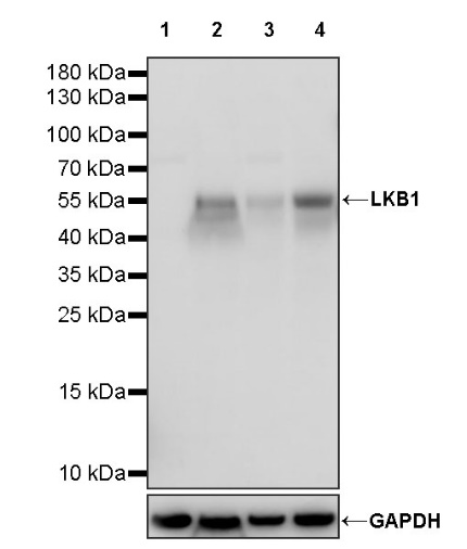
WB result of LKB1 Recombinant Rabbit mAb
Primary antibody: LKB1 Recombinant Rabbit mAb at 1/1000 dilution
Lane 1: HeLa whole cell lysate 20 µg
Lane 2: K562 whole cell lysate 20 µg
Lane 3: PANC-1 whole cell lysate 20 µg
Lane 4: 293T whole cell lysate 20 µg
Negative control: HeLa whole cell lysate
Secondary antibody: Goat Anti-rabbit IgG, (H+L), HRP conjugated at 1/10000 dilution
Predicted MW: 49 kDa
Observed MW: 55 kDa
WB result of LKB1 Recombinant Rabbit mAb
Primary antibody: LKB1 Recombinant Rabbit mAb at 1/1000 dilution
Lane 1: rat testis lysate 20 µg
Secondary antibody: Goat Anti-rabbit IgG, (H+L), HRP conjugated at 1/10000 dilution
Predicted MW: 49 kDa
Observed MW: 48, 55 kDa
Flow cytometric analysis of 4% PFA fixed 90% methanol permeabilized K562 (Human chronic myelogenous leukemia lymphoblast) labelling LKB1 antibody at 1/50 dilution (1 μg)/ (Red) compared with a Rabbit monoclonal IgG (Black) isotype control and an unlabelled control (cells without incubation with primary antibody and secondary antibody) (Blue). Goat Anti - Rabbit IgG Alexa Fluor® 488 was used as the secondary antibody.
LKB1 Rabbit mAb at 1/50 dilution (1 µg) immunoprecipitating LKB1 in 0.4 mg rat testis lysate.
Western blot was performed on the immunoprecipitate using LKB1 Rabbit mAb at 1/1000 dilution.
Secondary antibody (HRP) for IP was used at 1/1000 dilution.
Lane 1: rat testis lysate 20 µg (Input)
Lane 2: LKB1 Rabbit mAb IP in rat testis lysate
Lane 3: Rabbit monoclonal IgG IP in rat testis lysate
Predicted MW: 49 kDa
Observed MW: 48, 55 kDa
IHC shows positive staining in paraffin-embedded human testis. Anti- LKB1 antibody was used at 1/500 dilution, followed by a HRP Polymer for Mouse & Rabbit IgG (ready to use). Counterstained with hematoxylin. Heat mediated antigen retrieval with Tris/EDTA buffer pH9.0 was performed before commencing with IHC staining protocol.
IHC shows positive staining in paraffin-embedded mouse testis. Anti- LKB1 antibody was used at 1/500 dilution, followed by a HRP Polymer for Mouse & Rabbit IgG (ready to use). Counterstained with hematoxylin. Heat mediated antigen retrieval with Tris/EDTA buffer pH9.0 was performed before commencing with IHC staining protocol.
IHC shows positive staining in paraffin-embedded rat testis. Anti- LKB1 antibody was used at 1/500 dilution, followed by a HRP Polymer for Mouse & Rabbit IgG (ready to use). Counterstained with hematoxylin. Heat mediated antigen retrieval with Tris/EDTA buffer pH9.0 was performed before commencing with IHC staining protocol.
ICC shows positive staining in K562 cells. Anti-LKB1 antibody was used at 1/500 dilution (Green) and incubated overnight at 4°C. Goat polyclonal Antibody to Rabbit IgG - H&L (Alexa Fluor® 488) was used as secondary antibody at 1/1000 dilution. The cells were fixed with 4% PFA and permeabilized with 0.1% PBS-Triton X-100. Nuclei were counterstained with DAPI (Blue). Counterstain with tubulin (Red).
 您现在的位置: