
PBS, pH7.4, 0.03% Proclin 300
12 months from date of receipt / reconstitution, 2 to 8 °C as supplied
| 应用 | 稀释度 |
|---|---|
| FCM | 1:2000 |
CD4 (cluster of differentiation 4) is a glycoprotein that serves as a co-receptor for the T-cell receptor (TCR). CD4 is found on the surface of immune cells such as helper T cells, monocytes, macrophages, and dendritic cells. CD4 is a co-receptor of the T cell receptor (TCR) and assists the latter in communicating with antigen-presenting cells. The TCR complex and CD4 bind to distinct regions of the antigen-presenting MHC class II molecule. The extracellular D1 domain of CD4 binds to the β2 region of MHC class II. The resulting close proximity between the TCR complex and CD4 allows the tyrosine kinase Lck bound to the cytoplasmic tail of CD4 to phosphorylate tyrosine residues of immunoreceptor tyrosine activation motifs (ITAMs) on the cytoplasmic domains of CD3 to amplify the signal generated by the TCR. CD4 continues to be expressed in most neoplasms derived from T helper cells. It is therefore possible to use CD4 immunohistochemistry on tissue biopsy samples to identify most forms of peripheral T cell lymphoma and related malignant conditions. The antigen has also been associated with a number of autoimmune diseases such as vitiligo and type I diabetes mellitus.
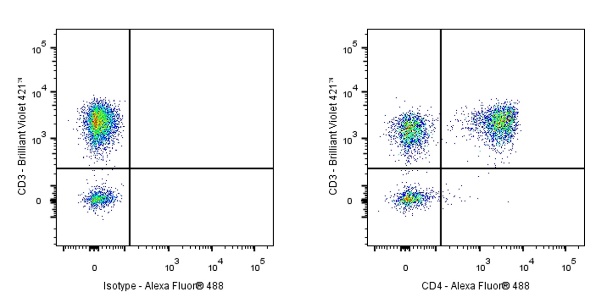
Flow cytometric analysis of human PBMC (human peripheral blood mononuclear cell) labelling CD4 Chimeric antibody at 1/2000 (0.1 μg) dilution (Right) compared with a Rabbit monoclonal IgG isotype control (Left). Goat Anti - Rabbit IgG Alexa Fluor® 488 was used as the secondary antibody. Then cells were stained with CD3 - Brilliant Violet 421™ separately. Gated on viable lymphocytes.
 您现在的位置: