
PBS, 40% Glycerol, 0.05% BSA, 0.03% Proclin 300
12 months from date of receipt / reconstitution, -20 °C as supplied
| 应用 | 稀释度 |
|---|---|
| WB | 1:1000 |
| IP | 1:50 |
| IHC-P | 1:500 |
| IF | 1:100 |
EGR2 (also termed Krox20) is a transcription regulatory factor, containing three zinc finger DNA-binding sites, and is highly expressed in a population of migrating neural crest cells. It is later expressed in the neural crest derived cells of the cranial ganglion. Mutations in this gene are associated with the autosomal dominant Charcot-Marie-Tooth disease, type 1D, Dejerine–Sottas disease, and Congenital Hypomyelinating Neuropathy.[18] Two studies have linked EGR2 expression to proliferation of osteoprogenitors and cell lines derived from Ewing sarcoma, which is a highly aggressive bone-associated cancer. New research suggests that EGR2 - or the lack of it - is the reason for male baldness.
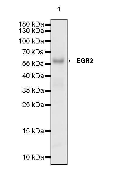
WB result of EGR2 Recombinant Rabbit mAb
Primary antibody: EGR2 Recombinant Rabbit mAb at 1/1000 dilution
Lane 1: mouse thymus lysate 20 µg
Secondary antibody: Goat Anti-rabbit IgG, (H+L), HRP conjugated at 1/10000 dilution
Predicted MW: 50 kDa
Observed MW: 58 kDa
EGR2 Rabbit mAb at 1/50 dilution (1 µg) immunoprecipitating EGR2 in 0.4 mg mouse thymus lysate.
Western blot was performed on the immunoprecipitate using EGR2 Rabbit mAb at 1/1000 dilution.
Secondary antibody (HRP) for IP was used at 1/1000 dilution.
Lane 1: mouse thymus lysate 20 µg (Input)
Lane 2: EGR2 Rabbit mAb IP in mouse thymus lysate
Lane 3: Rabbit monoclonal IgG IP in 2mouse thymus lysate
Predicted MW: 50 kDa
Observed MW: 58 kDa
This blot was developed with high sensitivity substrate
IHC shows positive staining in paraffin-embedded mouse cerebral cortex. Anti-EGR2 antibody was used at 1/500 dilution, followed by a HRP Polymer for Mouse & Rabbit IgG (ready to use). Counterstained with hematoxylin. Heat mediated antigen retrieval with Tris/EDTA buffer pH9.0 was performed before commencing with IHC staining protocol.
Negative control: IHC shows negative staining in paraffin-embedded mouse colon. Anti-EGR2 antibody was used at 1/500 dilution, followed by a HRP Polymer for Mouse & Rabbit IgG (ready to use). Counterstained with hematoxylin. Heat mediated antigen retrieval with Tris/EDTA buffer pH9.0 was performed before commencing with IHC staining protocol.
Negative control: IHC shows negative staining in paraffin-embedded mouse liver. Anti-EGR2 antibody was used at 1/500 dilution, followed by a HRP Polymer for Mouse & Rabbit IgG (ready to use). Counterstained with hematoxylin. Heat mediated antigen retrieval with Tris/EDTA buffer pH9.0 was performed before commencing with IHC staining protocol.
IHC shows positive staining in paraffin-embedded mouse spleen. Anti-EGR2 antibody was used at 1/500 dilution, followed by a HRP Polymer for Mouse & Rabbit IgG (ready to use). Counterstained with hematoxylin. Heat mediated antigen retrieval with Tris/EDTA buffer pH9.0 was performed before commencing with IHC staining protocol.
IHC shows positive staining in paraffin-embedded mouse thymus. Anti-EGR2 antibody was used at 1/500 dilution, followed by a HRP Polymer for Mouse & Rabbit IgG (ready to use). Counterstained with hematoxylin. Heat mediated antigen retrieval with Tris/EDTA buffer pH9.0 was performed before commencing with IHC staining protocol.
IHC shows positive staining in paraffin-embedded mouse skin. Anti-EGR2 antibody was used at 1/500 dilution, followed by a HRP Polymer for Mouse & Rabbit IgG (ready to use). Counterstained with hematoxylin. Heat mediated antigen retrieval with Tris/EDTA buffer pH9.0 was performed before commencing with IHC staining protocol.
IHC shows positive staining in paraffin-embedded rat cerebral cortex. Anti-EGR2 antibody was used at 1/500 dilution, followed by a HRP Polymer for Mouse & Rabbit IgG (ready to use). Counterstained with hematoxylin. Heat mediated antigen retrieval with Tris/EDTA buffer pH9.0 was performed before commencing with IHC staining protocol.
IF shows positive staining in paraffin-embedded mouse brain. Anti-EGR2 antibody was used at 1/100 dilution (Green) and incubated overnight at 4°C. Goat polyclonal Antibody to Rabbit IgG - H&L (Alexa Fluor® 488) was used as secondary antibody at 1/1000 dilution. Counterstained with DAPI (Blue). Heat mediated antigen retrieval with EDTA buffer pH9.0 was performed before commencing with IF staining protocol.
 您现在的位置: