
PBS, 40% Glycerol, 0.05% BSA, 0.03% Proclin 300
12 months from date of receipt / reconstitution, -20 °C as supplied
| 应用 | 稀释度 |
|---|---|
| WB | 1:1000 |
| IP | 1:50 |
PKC eta, also known as Protein Kinase C eta, is a member of the Protein Kinase C (PKC) family of enzymes. This serine/threonine-specific protein kinase plays crucial roles in various cellular processes, particularly in signal transduction, cell differentiation, and gene transcription regulation. PKC eta regulates keratinocyte differentiation by activating the MAPK13 (p38delta) activated protein kinase cascade. PKC eta mediates transcriptional activation of the transglutaminase 1 (TGM1) gene. Mutations in the gene have been linked to susceptibility to cerebral infarction.
WB result of PKC eta Recombinant Rabbit mAb
Primary antibody: PKC eta Recombinant Rabbit mAb at 1/1000 dilution
Lane 1: MCF7 whole cell lysate 20 µg
Lane 2: PC-3 whole cell lysate 20 µg
Secondary antibody: Goat Anti-rabbit IgG, (H+L), HRP conjugated at 1/10000 dilution
Predicted MW: 78 kDa
Observed MW: 45, 78 kDa
This blot was developed with high sensitivity substrate
WB result of PKC eta Recombinant Rabbit mAb
Primary antibody: PKC eta Recombinant Rabbit mAb at 1/1000 dilution
Lane 1: mouse lung lysate 20 µg
Secondary antibody: Goat Anti-rabbit IgG, (H+L), HRP conjugated at 1/10000 dilution
Predicted MW: 78 kDa
Observed MW: 45, 78 kDa
This blot was developed with high sensitivity substrate
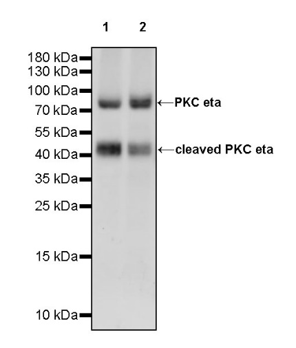
WB result of PKC eta Recombinant Rabbit mAb
Primary antibody: PKC eta Recombinant Rabbit mAb at 1/1000 dilution
Lane 1: rat lung lysate 20 µg
Lane 2: rat skin lysate 20 µg
Secondary antibody: Goat Anti-rabbit IgG, (H+L), HRP conjugated at 1/10000 dilution
Predicted MW: 78 kDa
Observed MW: 45, 78 kDa
This blot was developed with high sensitivity substrate
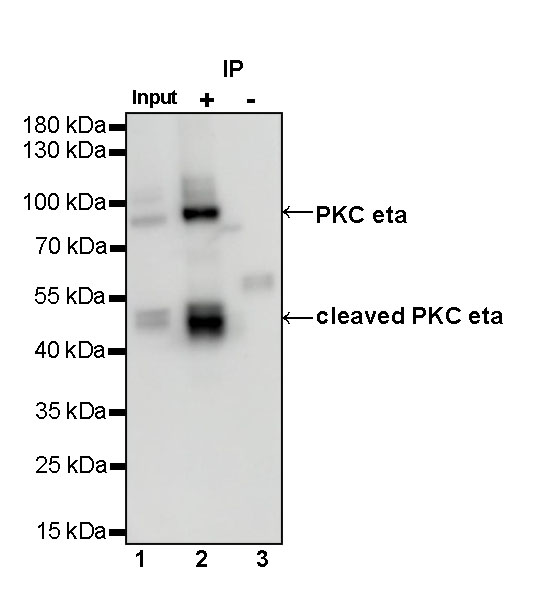
PKC eta Rabbit mAb at 1/50 dilution (1 µg) immunoprecipitating PKC eta in 0.4 mg mouse lung lysate.
Western blot was performed on the immunoprecipitate using PKC eta Rabbit mAb at 1/1000 dilution.
Secondary antibody (HRP) for IP was used at 1/1000 dilution.
Lane 1: mouse lung lysate 20 µg (Input)
Lane 2: PKC eta Rabbit mAb IP in mouse lung lysate
Lane 3: Rabbit monoclonal IgG IP in mouse lung lysate
Predicted MW: 78 kDa
Observed MW: 45, 78 kDa
 您现在的位置: