
PBS pH7.4, containing no preservative
2 to 8 °C for 2 weeks under sterile conditions;
-20 °C for 3 months under sterile conditions;
-80 °C for 24 months under sterile conditions.
Please avoid repeated freeze-thaw cycles.
| 应用 | 稀释度 |
|---|---|
| WB | 1:1000 |
| ICFCM | 1:200 |
| FCM | 1:200 |
Isotype control antibodies, to estimate the nonspecific binding of target.
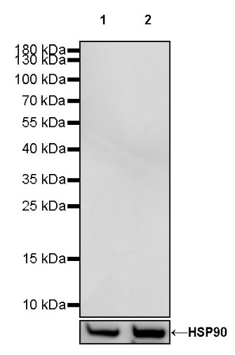
WB result of Invivo mouse IgG1 isotype control (D265A)
Primary antibody: Invivo mouse IgG1 isotype control (D265A) at 1/1000 dilution
Lane 1: HeLa whole cell lysate 20 µg
Lane 2: THP-1 whole cell lysate 20 µg
Secondary antibody: Goat Anti-mouse IgG, (H+L), HRP conjugated at 1/10000 dilution
WB result of Invivo mouse IgG1 isotype control (D265A)
Primary antibody: Invivo mouse IgG1 isotype control (D265A) at 1/1000 dilution
Lane 1: mouse brain lysate 20 µg
Secondary antibody: Goat Anti-mouse IgG, (H+L), HRP conjugated at 1/10000 dilution
WB result of Invivo mouse IgG1 isotype control (D265A)
Primary antibody: Invivo mouse IgG1 isotype control (D265A) at 1/1000 dilution
Lane 1: rat brain lysate 20 µg
Secondary antibody: Goat Anti-mouse IgG, (H+L), HRP conjugated at 1/10000 dilution
Flow cytometric analysis of THP-1 (Human monocytic leukemia monocyte) labeling mouse IgG1 isotype control (D265A) at 1/200 (1 μg) dilution (Left) / (Red) compared with CD13 antibody at 1/200 (1 μg) dilution (S0B0441) (Right) / (Red), Mouse monoclonal IgG Isotype Control (Left) / (Black) compared with mouse IgG1 isotype control (Right) / (Black) and an unlabelled control (cells without incubation with primary antibody and secondary antibody) (Blue). Goat Anti - Mouse IgG Alexa Fluor® 488 was used as the secondary antibody.
Flow cytometric analysis of 4% paraformaldehyde fixed 90% methanol permeabilized NIH/3T3 (Mouse embryonic fibroblast) labelling mouse IgG1 isotype control (D265A) at 1/200 (1 μg) dilution (Left) / (Red) compared with α-tubulin antibody (Right) / (Red), Mouse monoclonal IgG Isotype Control (Left) / (Black) compared with mouse IgG1 isotype control (Right) / (Black) and an unlabelled control (cells without incubation with primary antibody and secondary antibody) (Blue). Goat Anti - Mouse IgG Alexa Fluor® 488 was used as the secondary antibody.
IHC shows negative staining in paraffin-embedded human kidney. Invivo mouse IgG1 isotype control (D265A) was used at 1/200 dilution, followed by a HRP Polymer for Mouse & Rabbit IgG (ready to use). Counterstained with hematoxylin. Heat mediated antigen retrieval with Tris/EDTA buffer pH9.0 was performed before commencing with IHC staining protocol.
IHC shows negative staining in paraffin-embedded human liver. Invivo mouse IgG1 isotype control (D265A) was used at 1/200 dilution, followed by a HRP Polymer for Mouse & Rabbit IgG (ready to use). Counterstained with hematoxylin. Heat mediated antigen retrieval with Tris/EDTA buffer pH9.0 was performed before commencing with IHC staining protocol.
IHC shows negative staining in paraffin-embedded mouse spleen. Invivo mouse IgG1 isotype control (D265A) was used at 1/200 dilution, followed by a HRP Polymer for Mouse & Rabbit IgG (ready to use). Counterstained with hematoxylin. Heat mediated antigen retrieval with Tris/EDTA buffer pH9.0 was performed before commencing with IHC staining protocol.
 您现在的位置: