
PBS, 40% Glycerol, 0.05% BSA, 0.03% Proclin 300
12 months from date of receipt / reconstitution, -20 °C as supplied
| 应用 | 稀释度 |
|---|---|
| WB | 1:1000 |
| IP | 1:50 |
| ICC | 1:500 |
| ICFCM | 1:50 |
CTCF is a zinc finger protein that binds to specific DNA sequences known as CTCF-binding sites. It functions as a transcriptional regulator, chromatin organizer, and enhancer blocker. CTCF can either activate or repress gene expression, depending on the context and interacting proteins. It can bind to promoters, enhancers, and silencers to regulate transcription. CTCF is involved in the formation of chromatin loops and topologically associating domains (TADs). These loops and domains help organize the genome into functional units and regulate gene expression. CTCF can block the interaction between enhancers and promoters, preventing enhancer-mediated gene activation. This function is important for maintaining tissue-specific gene expression patterns. Mutations or abnormal expression of CTCF have been linked to various diseases, including cancer, developmental disorders, and neurological diseases.
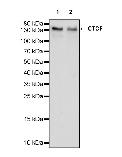
WB result of CTCF Recombinant Rabbit mAb
Primary antibody: CTCF Recombinant mAb at 1/1000 dilution
Lane 1: HCT 116 whole cell lysate 20 µg
Lane 2: HeLa whole cell lysate 20 µg
Secondary antibody: Goat Anti-rabbit IgG, (H+L), HRP conjugated at 1/10000 dilution
Predicted MW: 83 kDa
Observed MW: 140 kDa
This blot was developed with high sensitivity substrate
WB result of CTCF Recombinant Rabbit mAb
Primary antibody: CTCF Recombinant mAb at 1/1000 dilution
Lane 1: NIH/3T3 whole cell lysate 20 µg
Secondary antibody: Goat Anti-rabbit IgG, (H+L), HRP conjugated at 1/10000 dilution
Predicted MW: 83 kDa
Observed MW: 140 kDa
This blot was developed with high sensitivity substrate
WB result of CTCF Recombinant Rabbit mAb
Primary antibody: CTCF Recombinant mAb at 1/1000 dilution
Lane 1: C6 whole cell lysate 20 µg
Secondary antibody: Goat Anti-rabbit IgG, (H+L), HRP conjugated at 1/10000 dilution
Predicted MW: 83 kDa
Observed MW: 140 kDa
This blot was developed with high sensitivity substrate
Flow cytometric analysis of 4% PFA fixed 90% methanol permeabilized HeLa (Human cervix adenocarcinoma epithelial cell) labelling CTCF antibody at 1/50 dilution (1 μg)/ (Red) compared with a Rabbit monoclonal IgG (Black) isotype control and an unlabelled control (cells without incubation with primary antibody and secondary antibody) (Blue). Goat Anti - Rabbit IgG Alexa Fluor® 488 was used as the secondary antibody.
Flow cytometric analysis of 4% PFA fixed 90% methanol permeabilized NIH/3T3 (Mouse embryonic fibroblast) labelling CTCF antibody at 1/50 dilution (1 μg)/ (Red) compared with a Rabbit monoclonal IgG (Black) isotype control and an unlabelled control (cells without incubation with primary antibody and secondary antibody) (Blue). Goat Anti - Rabbit IgG Alexa Fluor® 488 was used as the secondary antibody.
CTCF Rabbit mAb at 1/50 dilution (1 µg) immunoprecipitating CTCF in 0.4 mg HeLa whole cell lysate.
Western blot was performed on the immunoprecipitate using CTCF Rabbit mAb at 1/1000 dilution.
Secondary antibody (HRP) for IP was used at 1/1000 dilution.
Lane 1: HeLa whole cell lysate 20 µg (Input)
Lane 2: CTCF Rabbit mAb IP in HeLa whole cell lysate
Lane 3: Rabbit monoclonal IgG IP in HeLa whole cell lysate
Predicted MW: 83 kDa
Observed MW: 140 kDa
ICC shows positive staining in HeLa cells. Anti-CTCF antibody was used at 1/500 dilution (Green) and incubated overnight at 4°C. Goat polyclonal Antibody to Rabbit IgG - H&L (Alexa Fluor® 488) was used as secondary antibody at 1/1000 dilution. The cells were fixed with 100% ice-cold methanol and permeabilized with 0.1% PBS-Triton X-100. Nuclei were counterstained with DAPI (Blue). Counterstain with tubulin (Red).
ICC shows positive staining in NIH/3T3 cells. Anti-CTCF antibody was used at 1/500 dilution (Green) and incubated overnight at 4°C. Goat polyclonal Antibody to Rabbit IgG - H&L (Alexa Fluor® 488) was used as secondary antibody at 1/1000 dilution. The cells were fixed with 4% PFA and permeabilized with 0.1% PBS-Triton X-100. Nuclei were counterstained with DAPI (Blue). Counterstain with tubulin (Red).
 您现在的位置: