
PBS, 40% Glycerol, 0.05% BSA, 0.03% Proclin 300
12 months from date of receipt / reconstitution, -20 °C as supplied
| 应用 | 稀释度 |
|---|---|
| WB | 1:1000 |
| IP | 1:50 |
| IHC-P | 1:500 |
UGGT1, known as UDP-Glucose Glycoprotein Glucosyltransferase 1, is an enzyme involved in the process of protein glycosylation. It is a soluble protein in the endoplasmic reticulum (ER) that selectively reglycosylates unfolded glycoproteins, providing quality control for protein trafficking out of the ER. In the process of hepatitis C virus (HCV) entry into cells, UGGT1 has been identified as a host factor that affects HCV entry by influencing SR-BI. As a biomarker, UGGT1 has potential applications in the diagnosis and treatment of cervical diseases such as cervical intraepithelial neoplasia and cervical squamous cell carcinoma.
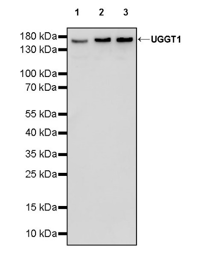
WB result of UGGT1 Recombinant Rabbit mAb
Primary antibody: UGGT1 Recombinant mAb at 1/1000 dilution
Lane 1: SH-SY5Y whole cell lysate 20 µg
Lane 2: Jurkat whole cell lysate 20 µg
Lane 3: MCF-7 whole cell lysate 20 µg
Secondary antibody: Goat Anti-rabbit IgG, (H+L), HRP conjugated at 1/10000 dilution
Predicted MW: 177 kDa
Observed MW: 170 kDa
UGGT1 Rabbit mAb at 1/50 dilution (1 µg) immunoprecipitating UGGT1 in 0.4 mg Jurkat whole cell lysate.
Western blot was performed on the immunoprecipitate using UGGT1 Rabbit mAb at 1/1000 dilution.
Secondary antibody (HRP) for IP was used at 1/1000 dilution.
Lane 1: Jurkat whole cell lysate 5 µg (Input)
Lane 2: UGGT1 Rabbit mAb IP in Jurkat whole cell lysate
Lane 3: Rabbit monoclonal IgG IP in Jurkat whole cell lysate
Predicted MW: 177 kDa
Observed MW: 170 kDa
This blot was developed with high sensitivity substrate
IHC shows positive staining in paraffin-embedded human cerebral cortex. Anti- UGGT1 antibody was used at 1/500 dilution, followed by a HRP Polymer for Mouse & Rabbit IgG (ready to use). Counterstained with hematoxylin. Heat mediated antigen retrieval with Tris/EDTA buffer pH9.0 was performed before commencing with IHC staining protocol.
IHC shows positive staining in paraffin-embedded human placenta. Anti- UGGT1 antibody was used at 1/500 dilution, followed by a HRP Polymer for Mouse & Rabbit IgG (ready to use). Counterstained with hematoxylin. Heat mediated antigen retrieval with Tris/EDTA buffer pH9.0 was performed before commencing with IHC staining protocol.
IHC shows positive staining in paraffin-embedded human gastric cancer. Anti- UGGT1 antibody was used at 1/500 dilution, followed by a HRP Polymer for Mouse & Rabbit IgG (ready to use). Counterstained with hematoxylin. Heat mediated antigen retrieval with Tris/EDTA buffer pH9.0 was performed before commencing with IHC staining protocol.
IHC shows positive staining in paraffin-embedded mouse cerebral cortex. Anti- UGGT1 antibody was used at 1/500 dilution, followed by a HRP Polymer for Mouse & Rabbit IgG (ready to use). Counterstained with hematoxylin. Heat mediated antigen retrieval with Tris/EDTA buffer pH9.0 was performed before commencing with IHC staining protocol.
IHC shows positive staining in paraffin-embedded rat cerebral cortex. Anti- UGGT1 antibody was used at 1/500 dilution, followed by a HRP Polymer for Mouse & Rabbit IgG (ready to use). Counterstained with hematoxylin. Heat mediated antigen retrieval with Tris/EDTA buffer pH9.0 was performed before commencing with IHC staining protocol.
 您现在的位置: