
12 months from date of receipt / reconstitution, -20 °C as supplied
| 应用 | 稀释度 |
|---|---|
| WB | 1:1000 |
| IHC-P | 1:250 |
| IP | 1:50 |
The VISTA protein, also known as V-domain immunoglobulin suppressor of T cell activation, plays a crucial role in immune regulation. The VISTA protein primarily functions as a T cell inhibitory ligand and receptor, participating in immune regulation by directly suppressing T cell function. As a co-inhibitory ligand, VISTA can act on yet undefined T cell-expressed receptors to inhibit the proliferation of CD4+ and CD8+ T cells, the production of IL-2 and IFN-γ, and promote the conversion of naive T cells into FoxP3+ regulatory T cells. As a co-inhibitory receptor, VISTA can suppress T cell function independently of antigen-presenting cells (APCs). VISTA-deficient mice exhibit spontaneous T cell activation, high levels of proinflammatory cytokine secretion, and more severe experimental autoimmune encephalomyelitis.
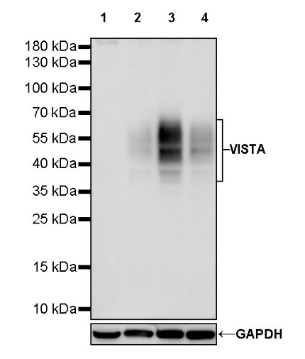
WB result of VISTA Recombinant Rabbit mAb
Primary antibody: VISTA Recombinant mAb at 1/1000 dilution
Lane 1: MCF-7 whole cell lysate 20 µg
Lane 2: HeLa whole cell lysate 20 µg
Lane 3: DU 145 whole cell lysate 20 µg
Lane 4: Jurkat whole cell lysate 20 µg
Negative control: MCF-7 whole cell lysate
Secondary antibody: Goat Anti-rabbit IgG, (H+L), HRP conjugated at 1/10000 dilution
Predicted MW: 34 kDa
Observed MW: 38~70 kDa
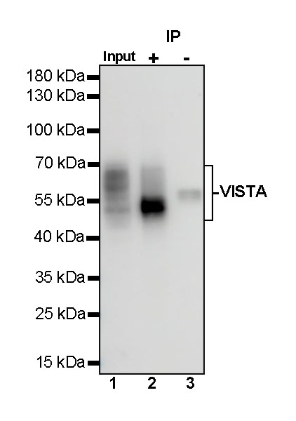
VISTA Rabbit mAb at 1/50 dilution (1 µg) immunoprecipitating VISTA in 0.4 mg DU 145 whole cell lysate.
Western blot was performed on the immunoprecipitate using VISTA Rabbit mAb at 1/1000 dilution.
Secondary antibody (HRP) for IP was used at 1/1000 dilution.
Lane 1: DU 145 whole cell lysate 20 µg (Input)
Lane 2: VISTA Rabbit mAb IP in DU 145 whole cell lysate
Lane 3: Rabbit monoclonal IgG IP in DU 145 whole cell lysate
Predicted MW: 34 kDa
Observed MW: 38~70 kDa
IHC shows positive staining in paraffin-embedded human cerebral cortex. Anti-VISAT antibody was used at 1/250 dilution, followed by a HRP Polymer for Mouse & Rabbit IgG (ready to use). Counterstained with hematoxylin. Heat mediated antigen retrieval with Tris/EDTA buffer pH9.0 was performed before commencing with IHC staining protocol.
IHC shows positive staining in paraffin-embedded human placenta. Anti-VISAT antibody was used at 1/250 dilution, followed by a HRP Polymer for Mouse & Rabbit IgG (ready to use). Counterstained with hematoxylin. Heat mediated antigen retrieval with Tris/EDTA buffer pH9.0 was performed before commencing with IHC staining protocol.
IHC shows positive staining in paraffin-embedded human cervical squamous cell carcinoma. Anti-VISAT antibody was used at 1/250 dilution, followed by a HRP Polymer for Mouse & Rabbit IgG (ready to use). Counterstained with hematoxylin. Heat mediated antigen retrieval with Tris/EDTA buffer pH9.0 was performed before commencing with IHC staining protocol.
IHC shows positive staining in paraffin-embedded human colon cancer. Anti-VISAT antibody was used at 1/250 dilution, followed by a HRP Polymer for Mouse & Rabbit IgG (ready to use). Counterstained with hematoxylin. Heat mediated antigen retrieval with Tris/EDTA buffer pH9.0 was performed before commencing with IHC staining protocol.
IHC shows positive staining in paraffin-embedded human lung adenocarcinoma. Anti-VISAT antibody was used at 1/250 dilution, followed by a HRP Polymer for Mouse & Rabbit IgG (ready to use). Counterstained with hematoxylin. Heat mediated antigen retrieval with Tris/EDTA buffer pH9.0 was performed before commencing with IHC staining protocol.
 您现在的位置: