
12 months from date of receipt / reconstitution, -20 °C as supplied
| 应用 | 稀释度 |
|---|---|
| FCM | 1:50 |
| ICC | 1:500 |
B7-H6, a novel member of the B7 family, has garnered significant attention in recent research. B7-H6 was identified in 2009 on the surface of tumor cells and recognized as a new member of the B7 family. B7-H6 has been implicated in tumorigenesis. It induces cytotoxicity, TNF-α, and IFN-γ secretion, promotes abnormal immune progression through HER2-scFv-mediated ADCC, and NKp30 immune evasion, thus facilitating tumor development. B7-H6 can inhibit apoptosis, promote tumor cell proliferation, and enhance the G0/G1 cell cycle process. It enhances the initiation of the "caspase cascade" and anti-apoptotic effects, and triggers tumorigenesis through STAT3 activation. B7-H6 is a ligand for NKp30, an activating receptor on natural killer (NK) cells. B7-H6 can activate NKp30-mediated NK cell activation, prompting cytokine secretion and exerting anti-tumor cytotoxic effects.
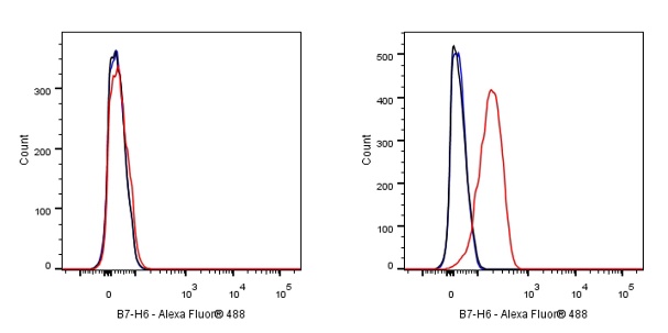
Flow cytometric analysis of MCF-7 (Human breast adenocarcinoma epithelial cell, left) / K562 (Human chronic myelogenous leukemia lymphoblast, right) cells labelling B7-H6 antibody at 1/50 dilution (1 μg) / (red) compared with a Rabbit monoclonal IgG (Black) isotype control and an unlabelled control (cells without incubation with primary antibody and secondary antibody) (Blue). Goat Anti - Rabbit IgG Alexa Fluor® 488 was used as the secondary antibody.
Negative control: MCF-7

ICC shows positive staining in K-562 cells (top panel) and negative staining in MCF7 cells (below panel). Anti-B7-H6 antibody was used at 1/500 dilution (Green) and incubated overnight at 4°C. Goat polyclonal Antibody to Rabbit IgG - H&L (Alexa Fluor® 488) was used as secondary antibody at 1/1000 dilution. The cells were fixed with 4% PFA and permeabilized with 0.1% PBS-Triton X-100. Nuclei were counterstained with DAPI (Blue). Counterstain with tubulin (Red).
 您现在的位置: