
12 months from date of receipt / reconstitution, -20 °C as supplied
| 应用 | 稀释度 |
|---|---|
| WB | 1:1000 |
| IHC-P | 1:2000 |
Cathepsin B is a crucial cysteine protease that plays a pivotal role in various physiological and pathological processes. Cathepsin B may enhance the activity of other proteases, including matrix metalloproteinase, urokinase (serine protease urokinase plasminogen activator), and cathepsin D, and thus it has an essential position for the proteolysis of extracellular matrix components, intercellular communication disruption, and reduced protease inhibitor expression. Cathepsin B is abnormally overexpressed in various malignancies, including lung cancer, gastric cancer, colon cancer, liver cancer, breast cancer, and prostate cancer. It degrades multiple extracellular matrix components, such as type I collagen, laminin, and proteoglycans, and activates interstitial procollagenase and type IV procollagenase, thereby participating in tumor invasion and metastasis. Studies have found that Cathepsin B exhibits higher activity under neutral or alkaline conditions in tumor tissues, which may be related to metabolic disorders in malignant tumors. Recent research has revealed a correlation between Cathepsin B and the pathogenesis of Alzheimer's disease (AD). Proteomic analysis of exosomes in the cerebrospinal fluid of AD patients showed that Cathepsin B and other proteins are associated with the onset and progression of AD.
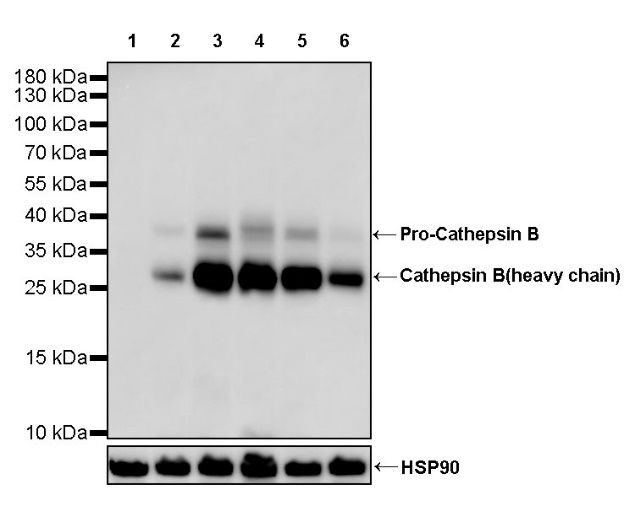
WB result of Cathepsin B Recombinant Rabbit mAb
Primary antibody: Cathepsin B Recombinant mAb at 1/1000 dilution
Lane 1: Daudi whole cell lysate 20 µg
Lane 2: A549 whole cell lysate 20 µg
Lane 3: SK-MEL-28 whole cell lysate 20 µg
Lane 4: HepG2 whole cell lysate 20 µg
Lane 5: HT-1080 whole cell lysate 20 µg
Lane 6: A375 whole cell lysate 20 µg
Negative control: Daudi whole cell lysate
Secondary antibody: Goat Anti-rabbit IgG, (H+L), HRP conjugated at 1/10000 dilution
Predicted MW: 38 kDa
Observed MW: 38, 28 kDa
IHC shows positive staining in paraffin-embedded human lung. Anti- Cathepsin B antibody was used at 1/2000 dilution, followed by a HRP Polymer for Mouse & Rabbit IgG (ready to use). Counterstained with hematoxylin. Heat mediated antigen retrieval with Tris/EDTA buffer pH9.0 was performed before commencing with IHC staining protocol.
IHC shows positive staining in paraffin-embedded human tonsil. Anti- Cathepsin B antibody was used at 1/2000 dilution, followed by a HRP Polymer for Mouse & Rabbit IgG (ready to use). Counterstained with hematoxylin. Heat mediated antigen retrieval with Tris/EDTA buffer pH9.0 was performed before commencing with IHC staining protocol.
IHC shows positive staining in paraffin-embedded human kidney. Anti- Cathepsin B antibody was used at 1/2000 dilution, followed by a HRP Polymer for Mouse & Rabbit IgG (ready to use). Counterstained with hematoxylin. Heat mediated antigen retrieval with Tris/EDTA buffer pH9.0 was performed before commencing with IHC staining protocol.
IHC shows positive staining in paraffin-embedded human cerebral cortex. Anti- Cathepsin B antibody was used at 1/2000 dilution, followed by a HRP Polymer for Mouse & Rabbit IgG (ready to use). Counterstained with hematoxylin. Heat mediated antigen retrieval with Tris/EDTA buffer pH9.0 was performed before commencing with IHC staining protocol.
IHC shows positive staining in paraffin-embedded human ovarian cancer. Anti- Cathepsin B antibody was used at 1/2000 dilution, followed by a HRP Polymer for Mouse & Rabbit IgG (ready to use). Counterstained with hematoxylin. Heat mediated antigen retrieval with Tris/EDTA buffer pH9.0 was performed before commencing with IHC staining protocol.
IHC shows positive staining in paraffin-embedded human lung squamous cell carcinoma. Anti- Cathepsin B antibody was used at 1/2000 dilution, followed by a HRP Polymer for Mouse & Rabbit IgG (ready to use). Counterstained with hematoxylin. Heat mediated antigen retrieval with Tris/EDTA buffer pH9.0 was performed before commencing with IHC staining protocol.
IHC shows positive staining in paraffin-embedded human cervical squamous cell carcinoma. Anti- Cathepsin B antibody was used at 1/2000 dilution, followed by a HRP Polymer for Mouse & Rabbit IgG (ready to use). Counterstained with hematoxylin. Heat mediated antigen retrieval with Tris/EDTA buffer pH9.0 was performed before commencing with IHC staining protocol.
 您现在的位置: