
12 months from date of receipt / reconstitution, -20 °C as supplied
| 应用 | 稀释度 |
|---|---|
| WB | 1:1000 |
| IHC-P | 1:500 |
| IP | 1:50 |
TFAM is a mitochondrial transcription factor that is a key activator of mitochondrial transcription as well as a participant in mitochondrial genome replication. TFAM binds mitochondrial promoter DNA to aid transcription of the mitochondrial genome. Studies in mice have demonstrated that this protein is required to regulate the mitochondrial genome copy number and is essential for embryonic development. A mouse model for Kearns–Sayre syndrome was produced when expression of this protein was eliminated by targeted disruption in heart and muscle cells.
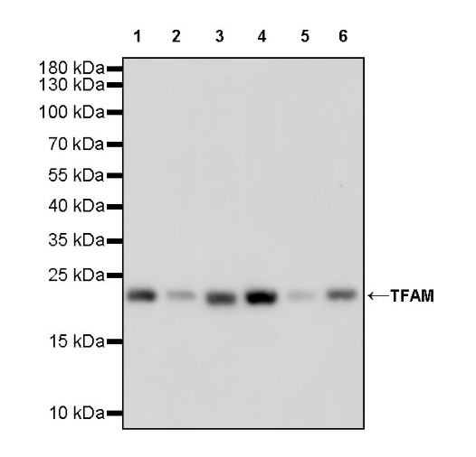
WB result of TFAM Recombinant Rabbit mAb
Primary antibody: TFAM Recombinant mAb at 1/1000 dilution
Lane 1: A431 whole cell lysate 20 µg
Lane 2: Jurkat whole cell lysate 20 µg
Lane 3: HepG2 whole cell lysate 20 µg
Lane 4: K562 whole cell lysate 20 µg
Lane 5: MCF7 whole cell lysate 20 µg
Lane 6: HEK-293 whole cell lysate 20 µg
Secondary antibody: Goat Anti-rabbit IgG, (H+L), HRP conjugated at 1/10000 dilution
Predicted MW: 29 kDa
Observed MW: 24 kDa
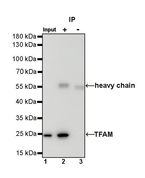
TFAM Rabbit mAb at 1/50 dilution (1 µg) immunoprecipitating TFAM in 0.4 mg A431 whole cell lysate.
Western blot was performed on the immunoprecipitate using TFAM Rabbit mAb at 1/1000 dilution.
Secondary antibody (HRP) for IP was used at 1/1000 dilution.
Lane 1: A431 whole cell lysate 20 µg (Input)
Lane 2: TFAM Rabbit mAb IP in A431 whole cell lysate
Lane 3: Rabbit monoclonal IgG IP in A431 whole cell lysate
Predicted MW: 29 kDa
Observed MW: 24 kDa
IHC shows positive staining in paraffin-embedded human testis. Anti-TFAM antibody was used at 1/500 dilution, followed by a HRP Polymer for Mouse & Rabbit IgG (ready to use). Counterstained with hematoxylin. Heat mediated antigen retrieval with Tris/EDTA buffer pH9.0 was performed before commencing with IHC staining protocol.
IHC shows positive staining in paraffin-embedded human kidney. Anti-TFAM antibody was used at 1/500 dilution, followed by a HRP Polymer for Mouse & Rabbit IgG (ready to use). Counterstained with hematoxylin. Heat mediated antigen retrieval with Tris/EDTA buffer pH9.0 was performed before commencing with IHC staining protocol.
IHC shows positive staining in paraffin-embedded human endometrial cancer. Anti-TFAM antibody was used at 1/500 dilution, followed by a HRP Polymer for Mouse & Rabbit IgG (ready to use). Counterstained with hematoxylin. Heat mediated antigen retrieval with Tris/EDTA buffer pH9.0 was performed before commencing with IHC staining protocol.
IHC shows positive staining in paraffin-embedded human colon cancer. Anti-TFAM antibody was used at 1/500 dilution, followed by a HRP Polymer for Mouse & Rabbit IgG (ready to use). Counterstained with hematoxylin. Heat mediated antigen retrieval with Tris/EDTA buffer pH9.0 was performed before commencing with IHC staining protocol.
IHC shows positive staining in paraffin-embedded human cervical squamous cell carcinoma. Anti-TFAM antibody was used at 1/500 dilution, followed by a HRP Polymer for Mouse & Rabbit IgG (ready to use). Counterstained with hematoxylin. Heat mediated antigen retrieval with Tris/EDTA buffer pH9.0 was performed before commencing with IHC staining protocol.
 您现在的位置: